. 2012 Oct;86(20):11066-77.
doi: 10.1128/JVI.01379-12. Epub 2012 Aug 1.

A mutation deleting sequences encoding the amino terminus of human cytomegalovirus UL84 impairs interaction with UL44 and capsid localization

Affiliations

A mutation deleting sequences encoding the amino terminus of human cytomegalovirus UL84 impairs interaction with UL44 and capsid localization

Blair L Strang et al. J Virol. 2012 Oct.

Abstract

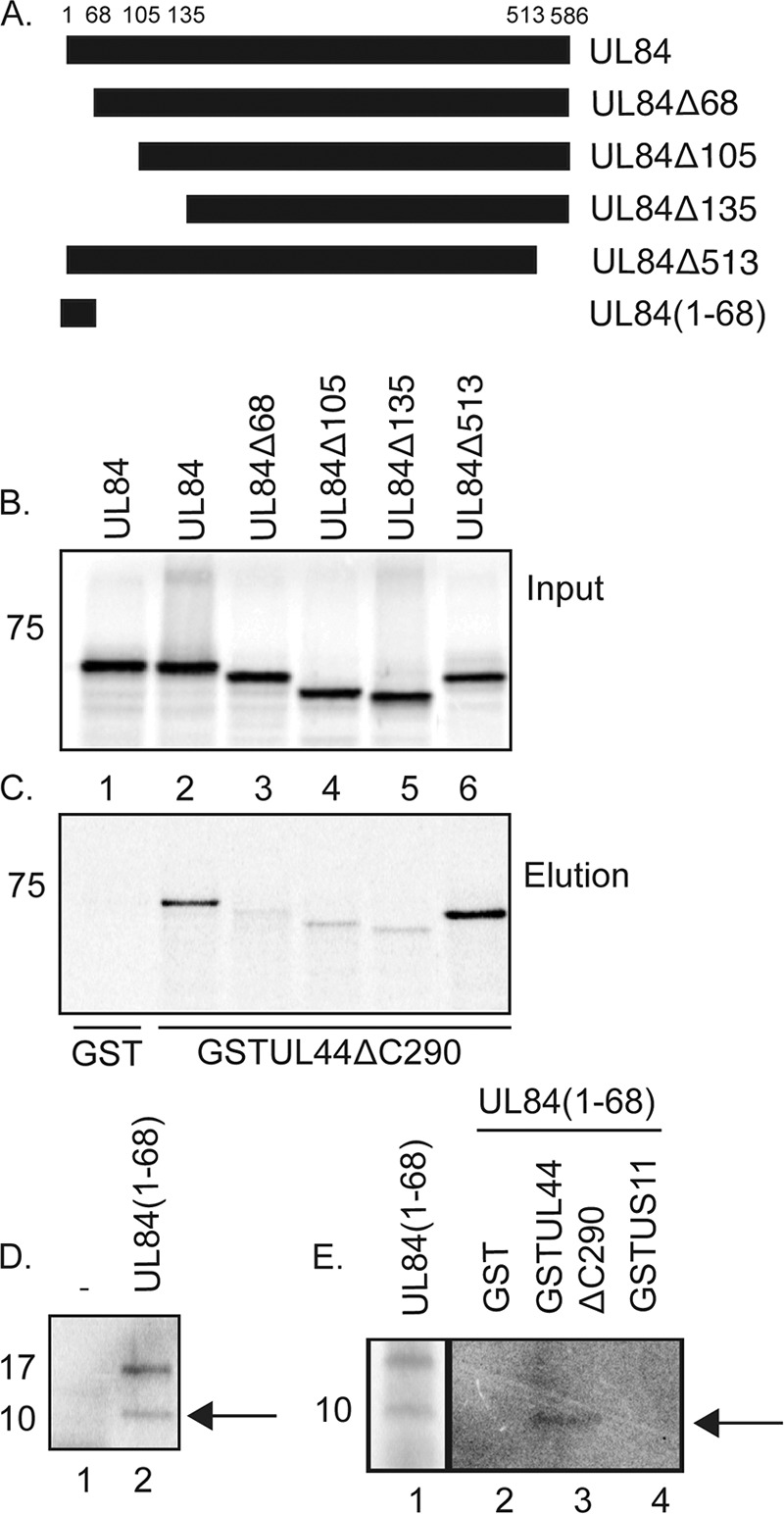
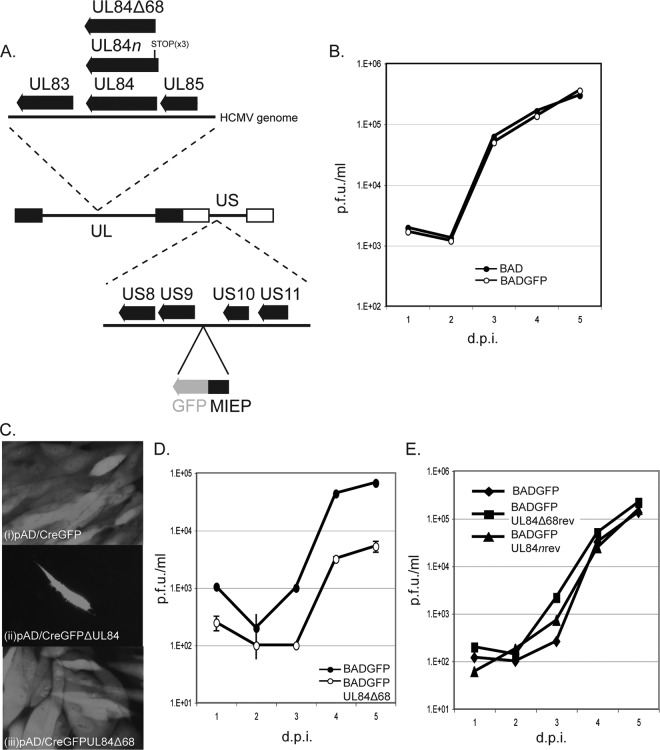
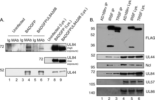
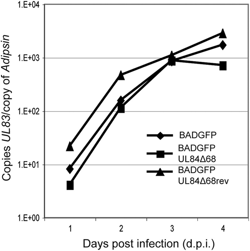
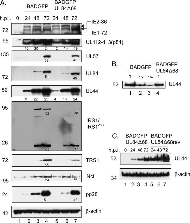
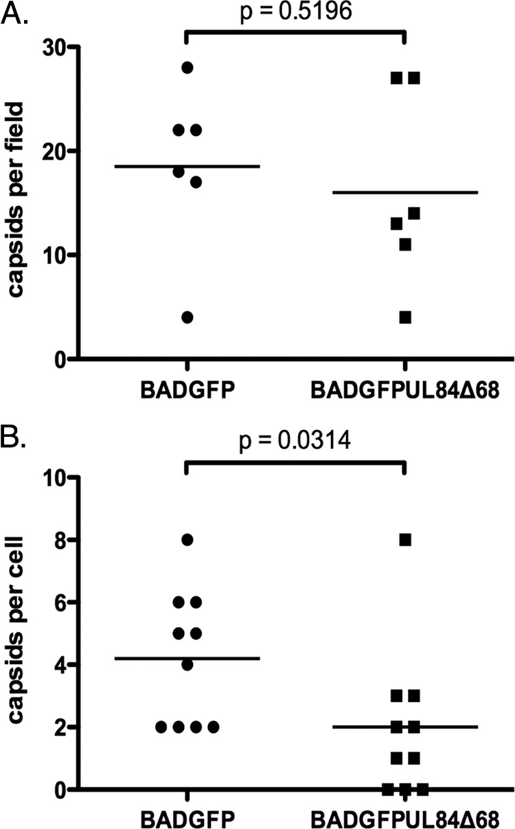
Protein-protein interactions are required for many biological functions. Previous work has demonstrated an interaction between the human cytomegalovirus DNA polymerase subunit UL44 and the viral replication factor UL84. In this study, glutathione S-transferase pulldown assays indicated that residues 1 to 68 of UL84 are both necessary and sufficient for efficient interaction of UL84 with UL44 in vitro. We created a mutant virus in which sequences encoding these residues were deleted. This mutant displayed decreased virus replication compared to wild-type virus. Immunoprecipitation assays showed that the mutation decreased but did not abrogate association of UL84 with UL44 in infected cell lysate, suggesting that the association in the infected cell can involve other protein-protein interactions. Further immunoprecipitation assays indicated that IRS1, TRS1, and nucleolin are candidates for such interactions in infected cells. Quantitative real-time PCR analysis of viral DNA indicated that the absence of the UL84 amino terminus does not notably affect viral DNA synthesis. Western blotting experiments and pulse labeling of infected cells with [(35)S]methionine demonstrated a rather modest downregulation of levels of multiple proteins and particularly decreased levels of the minor capsid protein UL85. Electron microscopy demonstrated that viral capsids assemble but are mislocalized in nuclei of cells infected with the mutant virus, with fewer cytoplasmic capsids detected. In sum, deletion of the sequences encoding the amino terminus of UL84 affects interaction with UL44 and virus replication unexpectedly, not viral DNA synthesis. Mislocalization of viral capsids in infected cell nuclei likely contributes to the observed decrease in virus replication.


Figures

Fig 1
Fig 1
Binding of UL84 and UL84 mutants to UL44 in vitro. (A) Schematic of UL84 and UL84 mutants used in GST pulldown assays. The residues of UL84 at which truncations have been made are noted above the figure. The name of each mutant is noted to the right of the figure. (B and C) GST pulldown assays were performed where GST or GST-UL44ΔC290 fusion proteins were incubated with radiolabeled UL84 or UL84 mutants shown in panel A produced by in vitro transcription/translation and passed over a glutathione column. The radiolabeled and GST proteins used in each reaction are noted above and below the figure, respectively. The input (B) and protein eluted by glutathione (C) from each reaction are shown. (D) Protein products of in vitro transcription/translation in reaction mixtures containing no protein expression vector (lane 1) or an expression vector encoding UL84(1–68) (lane 2) (from panel A). (E) GST pulldown assays were performed where GST, GST-UL44ΔC290, or GST-US11 fusion protein was incubated with radiolabeled UL84(1–68) and passed over a glutathione column. The GST proteins used in each reaction mixture are noted above the figure. Lane 1, input protein; lanes 2 to 4, protein eluted by glutathione from each reaction. The position of the approximately 10-kDa form of UL84(1–68) is indicated with an arrow in panels D and E. The position of molecular mass markers in panels B to E are indicated to the left of the figure.
Fig 2
Fig 2
Generation and characterization of virus expressing mutant UL84 proteins. (A) A schematic of the HCMV genome encoded in bacmid BADGFP is shown in the center of the figure. The unique long (UL) and unique short (US) segments of the genome are indicated. The internal and terminal repeats of UL and US are shown as black and white boxes, respectively. Dotted lines lead to magnified regions of the HCMV genome. Indicated below the HCMV genome is the intergenic region between open reading frames encoding US9 and US10 in which a cassette containing sequence encoding GFP (in gray) under the control of the major immediate-early promoter (MIEP) has been inserted. Indicated above the HCMV schematic of the genome is the region that contains the UL84 open reading frame. The insertion of stop codons into the UL84 open reading frame to create BADGFPUL84n is indicated with a vertical line. Also indicated is the truncation that creates BADGFPUL84Δ68. (B, D, and E) Viral replication. HFF cells were infected at an MOI of 1 with the indicated viruses. Virus supernatant was harvested at the indicated day postinfection (dpi). Virus titer is represented as PFU/ml (PFU/ml) on HFF cells. In each panel each data point represents the mean value from two independent experiments. In panel D, the error bars represent the standard error of the mean of the values used to calculate the data points. The titers of the virus stocks used in the assay were measured at the time the assays were performed to ensure that the correct inocula were used in each case. (C) Green fluorescence in cells electroporated with the indicated bacmid. Images are presented in grayscale.
Fig 3
Fig 3
IP of UL44 and UL84 from infected cell lysate. (A) Lysates from uninfected HFF cells or HFF cells infected with the indicated viruses (MOI of 3) were prepared and precleared with immunoglobulin. IP was then carried out with a monoclonal antibody (MAb) recognizing UL44 or a control antibody of the same isotype as the MAb used (Ig). Immunoprecipitated proteins were analyzed by Western blotting using MAb recognizing UL84 or UL44, as indicated to the right of the figure. Lanes 1 and 2, uninfected cells immunoprecipitated with Ig and MAb, respectively; lanes 3 and 4, BADGFP-infected cells immunoprecipitated with Ig and MAb, respectively; lanes 5 and 6, BADGFPUL84Δ68-infected cells immunoprecipitated with Ig and MAb, respectively; lanes 7 to 9, uninfected and infected cell lysate. Where indicated, high and low exposures of blot to film are shown. (B) Lysate from cells infected with BAD (lane 4), HCMV-IRSF (IRSF) (lane 5), and HCMV-TRSF (TRSF) (lane 6) and proteins immunoprecipitated using an anti-FLAG antibody from those lysates (lanes 1, 2, and 3, respectively) were separated on a 10% polyacrylamide gel. Proteins in each lane were examined by Western blotting for the presence of FLAG-tagged IRS1, FLAG-tagged TRS1, UL44, UL84, UL86, UL57, and nucleolin (Ncl) using antibodies recognizing these proteins, as indicated to the right of the figure. The positions of molecular weight mass (kDa) are indicated to the left of each figure.
Fig 4
Fig 4
Levels of viral DNA synthesis in infected cells. Viral DNA synthesis in each experiment was determined by quantitative real-time PCR at each of the time points indicated. The amount of viral DNA assayed is represented as copies of the viral gene UL83 per copy of the cellular adipsin gene.
Fig 5
Fig 5
Levels of viral and cellular proteins in infected cells. (A and C) Western blotting of infected cell lysate. HFF cells were infected at an MOI of 1 with the indicated viruses, and cell lysates were prepared for Western blotting at the indicated time points (indicated above the figure). (B) Determination of relative protein levels in cells. A 2-fold dilution series of protein from lane 2 of panel A (lanes 1 to 3) was analyzed by Western blotting using antibodies recognizing UL44 compared to undiluted protein from lane 5 of panel A. Proteins recognized by the antibodies used in each experiment are indicated to the right of each figure. The positions of molecular mass markers (kDa) are indicated to the left of each figure. In some panels the numbers below bands represent the percent adjusted volume of the signal from the particular bands measured within that panel using Quantity One software.
Fig 6
Fig 6
Levels of capsid proteins in infected cells. (A) Schematic of locus in the HCMV virus genome from which UL83 to UL85 are produced. Filled arrows above the genome represent proteins encoded within the genome. Black lines below the genome represent transcripts from which those proteins are produced. pA, polyadenylation signal. (B) Western blotting of infected cell lysate. HFF cells were infected at an MOI of 1 with the indicated viruses, and cell lysates were prepared for Western blotting at the indicated time points (indicated above the figure). (C) Determination of relative protein levels in cells. A 2-fold dilution series of protein from lane 4 of panel B (lanes 1 to 3) was analyzed by Western blotting compared to undiluted protein from lane 7 of panel B (lane 4). Proteins recognized by the antibodies used in each experiment are indicated to the right of each figure. The positions of molecular mass markers (kDa) are indicated to the left of each figure.
Fig 7
Fig 7
Electron microscopy of infected cells. Cells were infected with either BADGFP or BADGFPUL84Δ68 (A) or BADGFPUL84Δ68rev (B) were prepared for analysis by electron microscopy at 72 hpi. Panels in the left-hand column show microscopy images. Each white dot in panels in the middle column indicates the positions of a capsid in panels in the left-hand column. The microscopy images in the right-hand column are enlarged areas of the images in the left-hand column. (C) Electron microscopy image of a BADGFP-infected cell nucleus containing a nuclear dense body (NDB). The magnification of each image is indicated below the figure. The white scale bar represents 500 nm. (D) Representative examples of A, B, and C capsids (frames i to iii, respectively). NM, nuclear membrane.
Fig 8
Fig 8
Capsids detected in infected cells. (A) The total numbers of A, B, and C capsids in the nuclei of infected cells were counted. An image of infected cell nuclei taken at a magnification of ×9,600 was chosen at random from each infected cell analyzed in two independent experiments. (B) The total number of enveloped and nonenveloped capsids in the entire cytoplasm of an infected cell was counted at a magnification of ×9,600. The result of a Mann-Whitney test applied in each experiment is shown in each figure.

References

    1. Adamo JE, Schroer J, Shenk T. 2004. Human cytomegalovirus TRS1 protein is required for efficient assembly of DNA-containing capsids. J. Virol. 78:10221–10229 - PMC - PubMed
    1. Ahn JH, Jang WJ, Hayward GS. 1999. The human cytomegalovirus IE2 and UL112-113 proteins accumulate in viral DNA replication compartments that initiate from the periphery of promyelocytic leukemia protein-associated nuclear bodies (PODs or ND10). J. Virol. 73:10458–10471 - PMC - PubMed
    1. Appleton BA, et al. 2006. Crystal structure of the cytomegalovirus DNA polymerase subunit UL44 in complex with the C terminus from the catalytic subunit. Differences in structure and function relative to unliganded UL44. J. Biol. Chem. 281:5224–5232 - PubMed
    1. Appleton BA, Loregian A, Filman DJ, Coen DM, Hogle JM. 2004. The cytomegalovirus DNA polymerase subunit UL44 forms a C clamp-shaped dimer. Mol. Cell 15:233–244 - PubMed
    1. Baldick CJ, Jr, Marchini A, Patterson CE, Shenk T. 1997. Human cytomegalovirus tegument protein pp71 (ppUL82) enhances the infectivity of viral DNA and accelerates the infectious cycle. J. Virol. 71:4400–4408 - PMC - PubMed

Publication types

MeSH terms

LinkOut - more resources