. 2012 Oct;86(20):11128-37.
doi: 10.1128/JVI.01700-12. Epub 2012 Aug 1.

Severe acute respiratory syndrome coronavirus nsp1 facilitates efficient propagation in cells through a specific translational shutoff of host mRNA

Affiliations

Severe acute respiratory syndrome coronavirus nsp1 facilitates efficient propagation in cells through a specific translational shutoff of host mRNA

Tomohisa Tanaka et al. J Virol. 2012 Oct.

Abstract

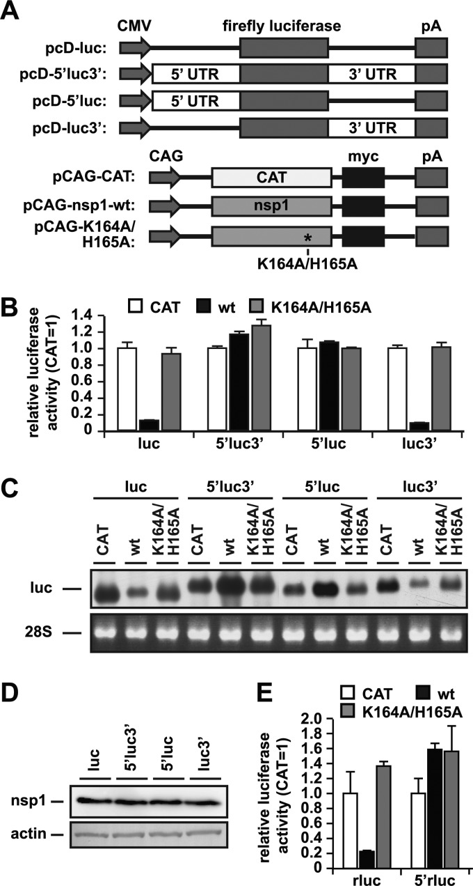
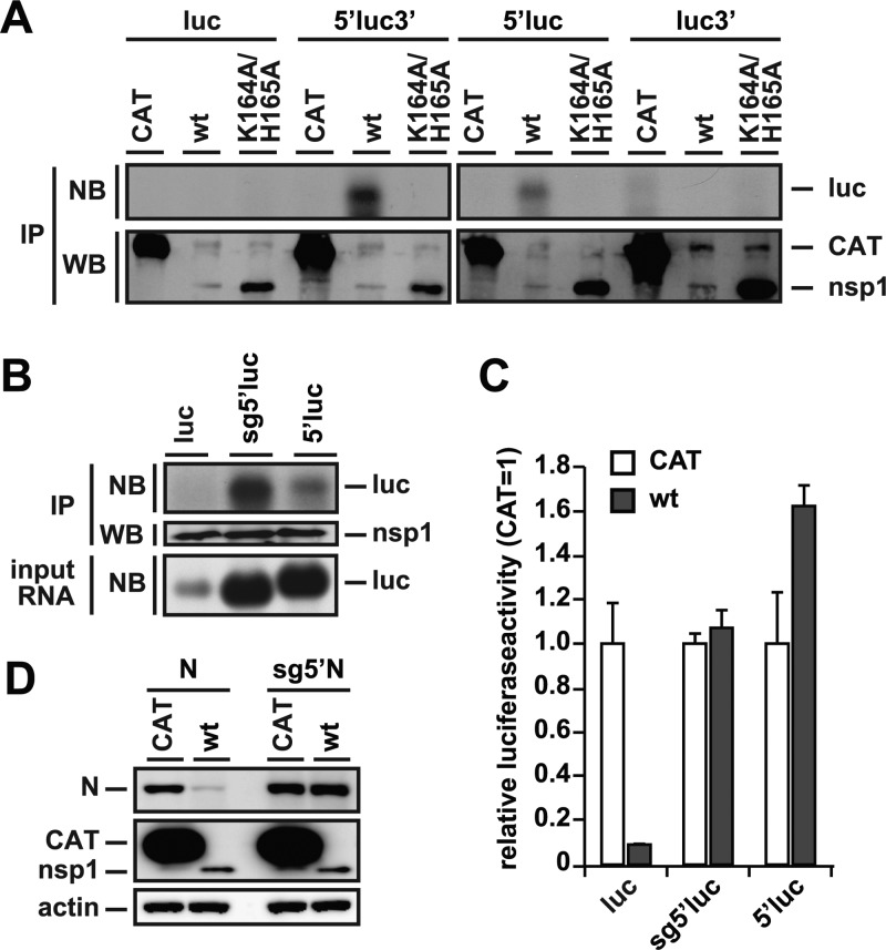
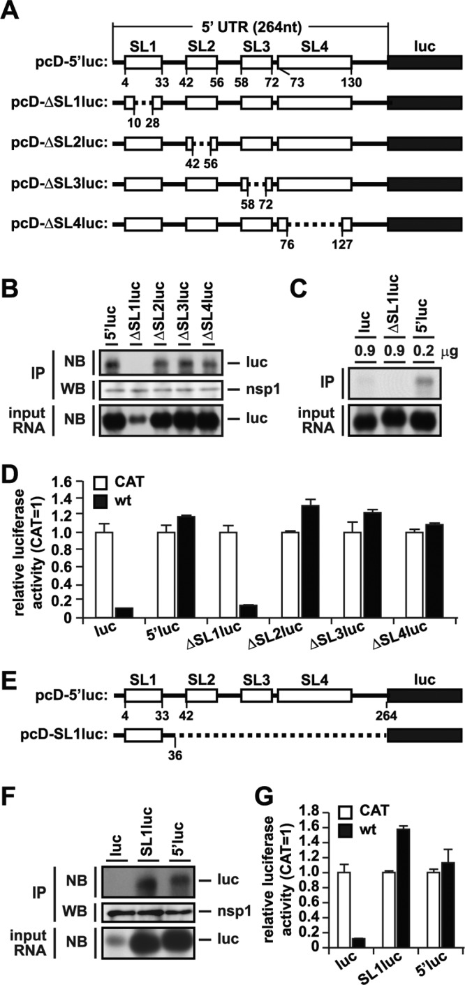
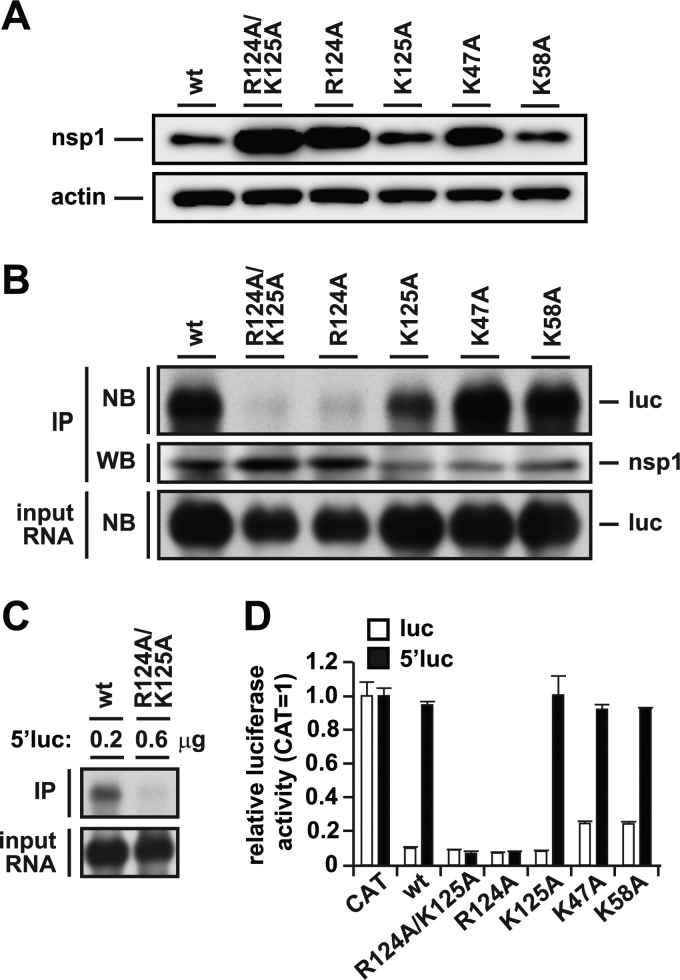
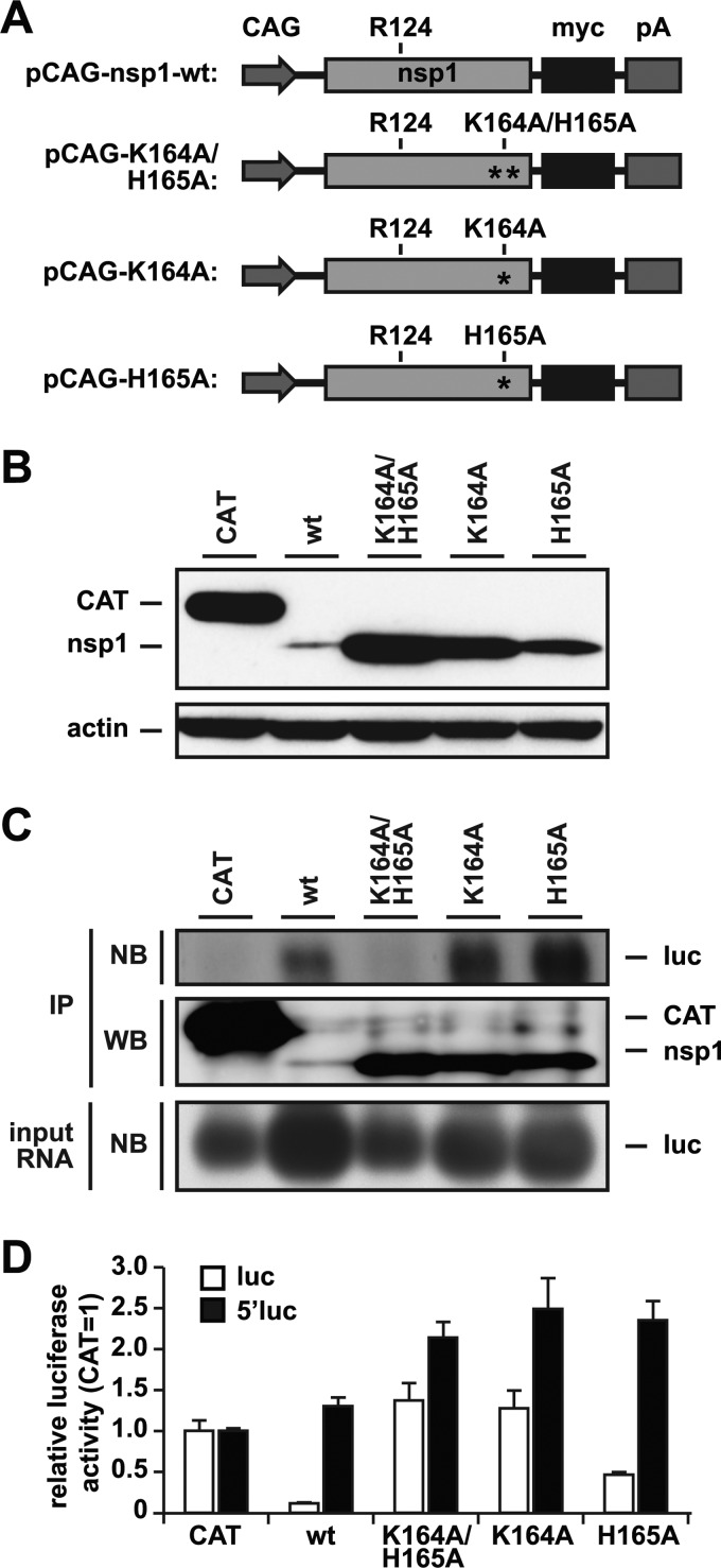
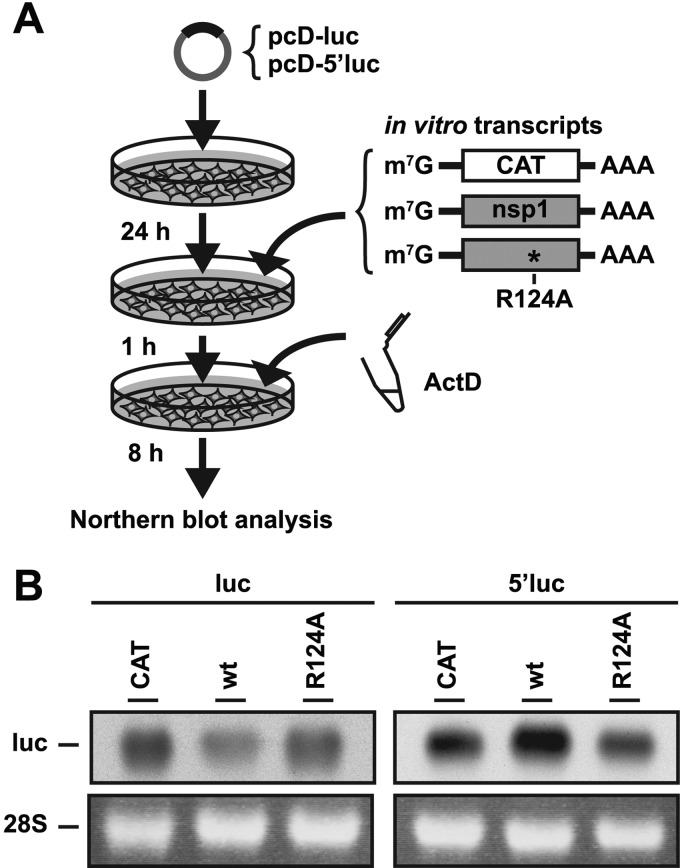
Severe acute respiratory syndrome (SARS) coronavirus (SCoV) is an enveloped virus containing a single-stranded, positive-sense RNA genome. Nine mRNAs carrying a set of common 5' and 3' untranslated regions (UTR) are synthesized from the incoming viral genomic RNA in cells infected with SCoV. A nonstructural SCoV nsp1 protein causes a severe translational shutoff by binding to the 40S ribosomal subunits. The nsp1-40S ribosome complex further induces an endonucleolytic cleavage near the 5'UTR of host mRNA. However, the mechanism by which SCoV viral proteins are efficiently produced in infected cells in which host protein synthesis is impaired by nsp1 is unknown. In this study, we investigated the role of the viral UTRs in evasion of the nsp1-mediated shutoff. Luciferase activities were significantly suppressed in cells expressing nsp1 together with the mRNA carrying a luciferase gene, while nsp1 failed to suppress luciferase activities of the mRNA flanked by the 5'UTR of SCoV. An RNA-protein binding assay and RNA decay assay revealed that nsp1 bound to stem-loop 1 (SL1) in the 5'UTR of SCoV RNA and that the specific interaction with nsp1 stabilized the mRNA carrying SL1. Furthermore, experiments using an SCoV replicon system showed that the specific interaction enhanced the SCoV replication. The specific interaction of nsp1 with SL1 is an important strategy to facilitate efficient viral gene expression in infected cells, in which nsp1 suppresses host gene expression. Our data indicate a novel mechanism of viral gene expression control by nsp1 and give new insight into understanding the pathogenesis of SARS.


Figures

Fig 1
Fig 1
The SCoV 5′UTR participates in evasion of nsp1-mediated shutoff and RNA degradation. (A) Schematic diagrams of the reporter plasmids carrying the firefly luciferase (luc) gene with or without the 5′UTR and/or 3′UTR of SCoV under the control of the CMV promoter and of those carrying the nsp1 or chloramphenicol acetyltransferase (CAT) gene under the control of the CAG promoter. pA, polyadenylation signal. The asterisk represents the mutated amino acid position. (B) Luciferase activities in 293T cells transfected with 1 μg of either pCAG-nsp1-wt (wt), pCAG-K164A/H165A (K164A/H165A), or pCAG-CAT (CAT) together with 0.2 μg of the indicated reporter plasmids were determined at 24 h posttransfection after standardization with those in cells expressing CAT. The values represent the means ± standard deviations (SD) from three independent experiments. (C) Total RNAs prepared from the cells as shown in panel B were subjected to Northern blot analysis by using a riboprobe for the luciferase gene (luc). 28S rRNA was stained with ethidium bromide (28S). (D) Lysates of the cells as described in panel B were subjected to Western blot analysis using anti-myc and antiactin antibodies. (E) Renilla luciferase activities in 293T cells transfected with 0.5 μg of either pCAG-nsp1-wt (wt), pCAG-K164A/H165A (K164A/H165A), or pCAG-CAT (CAT) together with 0.1 μg of the indicated reporter plasmids encoding Renilla luciferase were determined at 24 h posttransfection after standardization with those in cells expressing CAT.
Fig 2
Fig 2
Specific interaction of nsp1 with the 5′UTR confers resistance to nsp1-mediated gene suppression. (A) Lysates of 293T cells transfected with 1 μg of either pCAG-nsp1-wt (wt), pCAG-K164A/H165A (K164A/H165A), or pCAG-CAT (CAT) together with 0.2 μg of the indicated reporter plasmids were immunoprecipitated with anti-myc antibody at 30 h posttransfection. RNAs extracted from the precipitates were subjected to Northern blot analysis using a riboprobe for the luciferase gene (NB). The precipitates were also subjected to Western blot analysis using anti-myc antibody (WB). (B) Lysates of 293T cells transfected with 1 μg of pCAG-nsp1-wt together with 0.2 μg of the indicated reporter plasmids were immunoprecipitated with anti-myc antibody at 30 h posttransfection. RNAs extracted from the precipitates were subjected to Northern blot analysis using a riboprobe for the luciferase gene (NB, top). The precipitates were also subjected to Western blot analysis using anti-myc antibody (WB). The bottom panel represents the amount of intracellular reporter RNAs in the lysates. (C) Luciferase activities in 293T cells transfected as described for panel B were determined at 24 h posttransfection after standardization with those in cells expressing CAT. The values represent the means ± SD from three independent experiments. (D) Lysates of 293T cells transfected with 1 μg of either pCAG-nsp1-wt (wt) or pCAG-CAT (CAT) together with 0.2 μg of either pcD-N (N) or pcD-sg5′N (sg5′N) were subjected to Western blot analysis by using either anti-N (top), anti-myc (middle), or anti-actin (bottom) antibody at 24 h posttransfection.
Fig 3
Fig 3
Interaction of SL1 in the 5′UTR of SCoV with nsp1 is crucial for evasion of nsp1-mediated translational suppression. (A) Schematic diagram of deletion mutants derived from the pcD-5′luc plasmid. White boxes, dashed lines, and black boxes indicate stem-loops (SL1, SL2, SL3, and SL4) in the 5′UTR, deletions, and the firefly luciferase gene (luc), respectively. The numbers indicate nucleotide positions. (B) Lysates of 293T cells transfected with 1 μg of pCAG-nsp1-wt together with 0.2 μg of the indicated reporter plasmids were immunoprecipitated with anti-myc antibody at 30 h posttransfection. RNAs extracted from the precipitates were subjected to Northern blot analysis using a riboprobe for the luciferase gene (NB, top). The precipitates were also subjected to Western blot analysis using anti-myc antibody (WB). The bottom panel represents the amount of intracellular reporter RNAs in the lysates. (C) 293T cells were transfected with 1 μg of pCAG-nsp1-wt together with either 0.9 μg of pcD-luc (luc), 0.9 μg of pcD-ΔSL1luc (ΔSL1luc), or 0.2 μg of pcD-5′luc (5′luc), and the cell extracts were immunoprecipitated by anti-myc antibody. RNAs extracted from the precipitates were subjected to Northern blot analysis using a riboprobe for the luciferase gene (top). The bottom panel represents the amount of intracellular reporter RNAs in the lysates. (D) Luciferase activities in 293T cells transfected as described for panel B were determined at 24 h posttransfection after standardization with those in cells expressing CAT. The values represent the means ± SD from three independent experiments. (E) Schematic diagram of the pcD-SL1luc plasmid. Symbols are the same as in panel A. (F) Lysates of 293T cells transfected with 0.5 μg of pCAG-nsp1-wt together with 0.1 μg of the indicated reporter plasmids were immunoprecipitated with anti-myc antibody at 30 h posttransfection. RNAs extracted from the precipitates were subjected to Northern blot analysis using a riboprobe for the luciferase gene (NB, top). The precipitates were also subjected to Western blot analysis using anti-myc antibody (WB). The bottom panel represents the amount of intracellular reporter RNAs in the lysates. (G) Luciferase activities in 293T cells transfected as described for panel F were determined at 24 h posttransfection after standardization with those in cells expressing CAT. The values represent the means ± SD from three independent experiments.
Fig 4
Fig 4
Interaction of the 5′UTR of SCoV with nsp1 through R124 is required for the evasion of the nsp1-mediated translational suppression. (A) Expression of nsp1 in 293T cells transfected with 1 μg of either pCAG-nsp1-wt (wt), pCAG-R124A/K125A (R124A/K125A), pCAG-R124A (R124A), pCAG-K125A (K125A), pCAG-K47A (K47A), or pCAG-K58A (K58A) was examined by Western blot analysis using anti-myc or anti-actin antibody at 24 h posttransfection. (B) Lysates of 293T cells transfected with 1 μg of each of the expression plasmids as described for panel A together with 0.2 μg of pcD-5′luc (5′luc) were immunoprecipitated with anti-myc antibody at 30 h posttransfection. RNAs extracted from the precipitates were subjected to Northern blot analysis using a riboprobe for the luciferase gene (NB, top). The precipitates were also subjected to Western blot analysis using anti-myc antibody (WB). The bottom panel represents the amount of intracellular reporter RNAs in the lysates. (C) 293T cells were transfected with 1 μg of either pCAG-nsp1-wt (wt) or pCAG-R124A/K125A (R124A/K125A) together with either 0.2 or 0.6 μg of pcD-5′luc, and cell extracts were immunoprecipitated with anti-myc antibody at 30 h posttransfection. RNAs extracted from the precipitates were subjected to Northern blot analysis using a riboprobe for the luciferase gene (top). The bottom panel represents the amount of intracellular reporter RNAs in the lysates. (D) Luciferase activities in 293T cells transfected with 0.5 μg of either expression plasmids as described for panel A or pCAG-CAT (CAT) together with 0.1 μg of either pcD-luc (luc) or pcD-5′luc (5′luc) were determined at 24 h posttransfection after standardization with those in cells expressing CAT. The values represent the means ± SD from three independent experiments.
Fig 5
Fig 5
Two functional domains in nsp1 participate in RNA binding and translational shutoff. (A) Schematic diagrams of pCAG-nsp1-wt, pCAG-K164A/H165A, pCAG-K164A, and pCAG-H165A. CAG, CAG promoter; pA, polyadenylation signal. Asterisks represent the mutated amino acid position. (B) Expression of nsp1 in 293T cells transfected with 1 μg of either pCAG-nsp1-wt (wt), pCAG-K164A/H165A (K164A/H165A), pCAG-K164A (K164A), pCAG-H165A (H165A), or pCAG-CAT (CAT) was examined by Western blot analysis using anti-myc or antiactin antibody at 24 h posttransfection. (C) Lysates of 293T cells transfected with 1 μg of expression plasmids as described for panel A together with 0.2 μg of pcD-5′luc (5′luc) were immunoprecipitated with anti-myc antibody at 30 h posttransfection. RNAs extracted from the precipitates were subjected to Northern blot analysis using a riboprobe for luc (NB, top). The precipitates were also subjected to Western blot analysis using anti-myc antibody (WB). The bottom panel represents the amount of intracellular reporter RNAs in the lysates. (D) Luciferase activities in 293T cells transfected with 0.5 μg of either expression plasmids as described for panel A together with 0.1 μg of either pcD-luc (luc) or pcD-5′luc (5′luc) were determined at 24 h posttransfection after standardization with those in cells expressing CAT. The values represent the means ± SD from three independent experiments.
Fig 6
Fig 6
Nsp1 enhances the stability of RNA carrying the 5′UTR of SCoV. (A) Scheme for the RNA decay assay. 293T cells transfected with 0.25 μg of either pcD-luc (luc) or pcD-5′luc (5′luc) were further transfected with 1 μg each of RNAs carrying cap and poly(A) in the 5′ and 3′ ends, respectively, encoding either nsp1-wt, R124A, or CAT fused with myc-His tag in the C terminus at 24 h after primary transfection. Cells were further treated with 4 μg/ml of actinomycin D (ActD) at 1 h after RNA transfection, and intracellular RNAs were extracted at 8 h posttreatment and subjected to Northern blot analysis using a riboprobe for the luciferase gene. (B) Amounts of the luciferase RNA were examined by using Northern blot analysis (upper panels). 28S rRNA was stained with ethidium bromide (lower panels).
Fig 7
Fig 7
Specific interaction of nsp1 with the 5′UTR is required for efficient replication of SCoV. (A) Schematic diagram of cDNAs of the RNA replicon of SCoV. CMV, CMV promoter; TRS, transcription-regulatory sequence of SCoV; replicase, replicase gene of SCoV; pA, synthetic poly(A) tail; Rz, the self-cleaving ribozyme of hepatitis delta virus; BGH, BGH termination and polyadenylation signal. Asterisks represent the position of mutated amino acids. (B) 293T cells were transfected with 2.2 μg of either pBAC-wt or pBAC-R124As and the expression of viral RNA and proteins was determined at 12, 24, and 36 h posttransfection. Total RNAs extracted from the cells were subjected to Northern blot analysis using a riboprobe for the N gene. 28S rRNA was stained with ethidium bromide. Expression of N, nsp1, and actin was determined by Western blot analysis by using anti-N, anti-nsp1, and anti-actin antibody, respectively. The asterisk indicates a nonspecific signal. (C) Luciferase activities in 293T cells transfected with 1 μg of either pBAC-wt-rluc or pBAC-R124A-rluc were determined at 12, 24, and 36 h posttransfection. The values represent the means ± SD from three independent experiments. (D and E) 293T cells were transfected with 2.2 μg of pBAC-R124A-rluc together with 1 μg of either pCAG-nsp1-wt (wt) or pCAG-R124A (R124A), and the expression of viral RNA and proteins was determined at 24 and 36 h posttransfection. Total RNAs extracted from the cells were subjected to Northern blot analysis using a riboprobe for the Renilla luciferase gene (rluc). 28S rRNA was stained with ethidium bromide. Expression of nsp1 and actin was determined by Western blot analysis by using anti-myc and antiactin antibody, respectively. Luciferase activities in the cells were determined at 24 and 36 h posttransfection. The values represent the means ± SD from three independent experiments.

Similar articles

Cited by

References

    1. Almazán F, et al. 2006. Construction of a severe acute respiratory syndrome coronavirus infectious cDNA clone and a replicon to study coronavirus RNA synthesis. J. Virol. 80:10900–10906 - PMC - PubMed
    1. Almeida MS, Johnson MA, Herrmann T, Geralt M, Wüthrich K. 2007. Novel beta-barrel fold in the nuclear magnetic resonance structure of the replicase nonstructural protein 1 from the severe acute respiratory syndrome coronavirus. J. Virol. 81:3151–3161 - PMC - PubMed
    1. Barretto N, et al. 2005. The papain-like protease of severe acute respiratory syndrome coronavirus has deubiquitinating activity. J. Virol. 79:15189–15198 - PMC - PubMed
    1. Cencic R, et al. 2011. Blocking eIF4E-eIF4G interaction as a strategy to impair coronavirus replication. J. Virol. 85:6381–6389 - PMC - PubMed
    1. Chen S, Olsthoorn R. 2010. Group-specific structural features of the 5′-proximal sequences of coronavirus genomic RNAs. Virology 401:29–41 - PMC - PubMed

Publication types

MeSH terms

LinkOut - more resources