. 2012 Sep;23(18):3694-706.
doi: 10.1091/mbc.E12-03-0206. Epub 2012 Aug 1.

SRSF1 regulates the assembly of pre-mRNA processing factors in nuclear speckles

Affiliations

SRSF1 regulates the assembly of pre-mRNA processing factors in nuclear speckles

Vidisha Tripathi et al. Mol Biol Cell. 2012 Sep.

Abstract

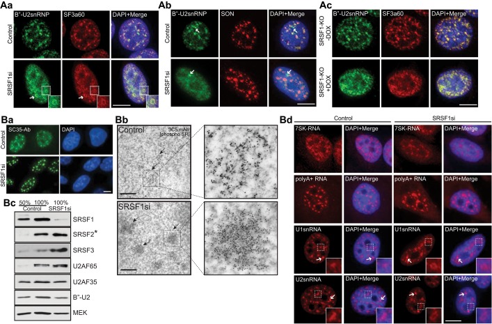
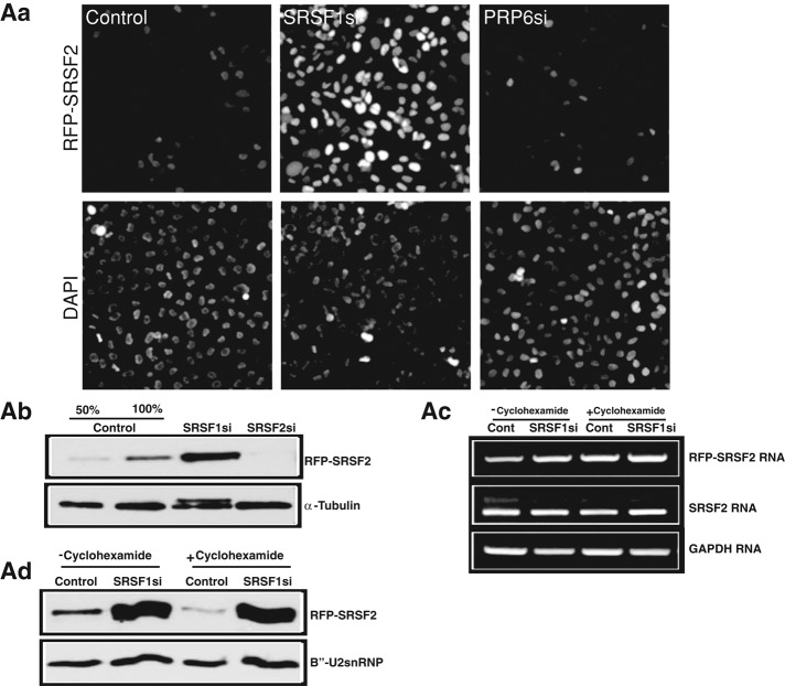
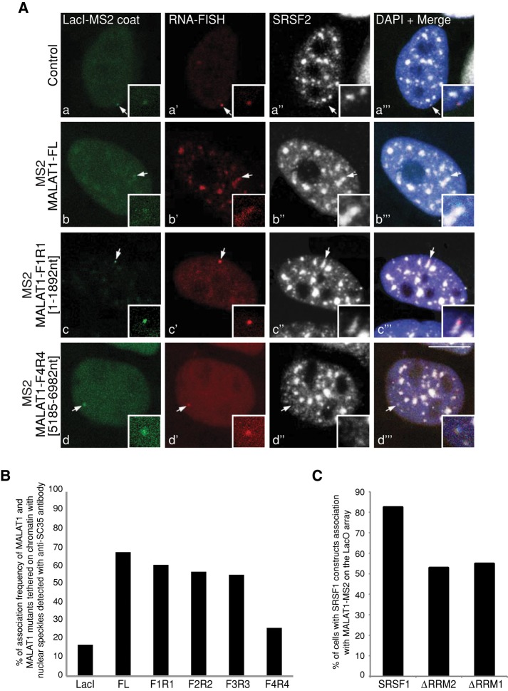
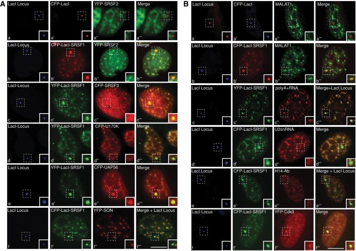
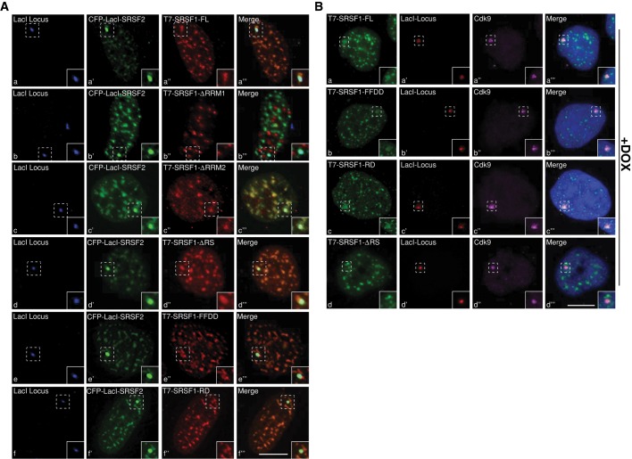
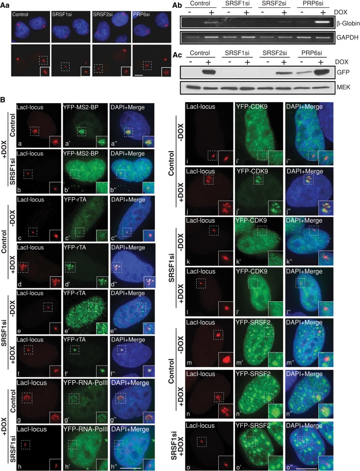
The mammalian cell nucleus is compartmentalized into nonmembranous subnuclear domains that regulate key nuclear functions. Nuclear speckles are subnuclear domains that contain pre-mRNA processing factors and noncoding RNAs. Many of the nuclear speckle constituents work in concert to coordinate multiple steps of gene expression, including transcription, pre-mRNA processing and mRNA transport. The mechanism that regulates the formation and maintenance of nuclear speckles in the interphase nucleus is poorly understood. In the present study, we provide evidence for the involvement of nuclear speckle resident proteins and RNA components in the organization of nuclear speckles. SR-family splicing factors and their binding partner, long noncoding metastasis-associated lung adenocarcinoma transcript 1 RNA, can nucleate the assembly of nuclear speckles in the interphase nucleus. Depletion of SRSF1 in human cells compromises the association of splicing factors to nuclear speckles and influences the levels and activity of other SR proteins. Furthermore, on a stably integrated reporter gene locus, we demonstrate the role of SRSF1 in RNA polymerase II-mediated transcription. Our results suggest that SR proteins mediate the assembly of nuclear speckles and regulate gene expression by influencing both transcriptional and posttranscriptional activities within the cell nucleus.


Figures

FIGURE 1:
FIGURE 1:
Depletion of SRSF1 results in the disorganization of nuclear speckle components. (A, a and b) Immunofluorescence staining using antibodies against B′′-U2snRNP, SF3a60, and SON in control and SRSF1 siRNA-treated HeLa cells. The arrows (A, b) designate Cajal bodies. (A, c) Immunofluorescence staining using antibodies against B′′-U2snRNP and SF3a60 in control and SRSF1-knockout MEFs. Immunofluorescence (B, a; SC35 antibody) and immune-EM (B, b; 3C5 antibody) analyses using antibodies that preferentially detect phosphorylated SR proteins in control and SRSF1-siRNA–treated HeLa cells. (B, c) Immunoblot analyses using antibodies against various pre-mRNA processing factors in control and SRSF1-depleted total cellular extracts. Asterisk, SRSF2 antibody preferentially recognizes the unphosphorylated or hypophosphorylated forms of SRSF2 (Bubulya et al., 2004). (B, d) RNA-FISH (red) of 7SK RNA, poly(A)+ RNA, and U1 and U2 snRNA in control and SRSF1-depleted HeLa cells. The DNA is counterstained with DAPI. Scale bars, fluorescent and EM images, 5 and 1 μm, respectively.
FIGURE 2:
FIGURE 2:
SRSF1-depletion results in the stabilization of the cellular pool of SRSF2. (A, a) SRSF1-depleted HeLa cells show increased cellular levels of RFP-SRSF2. (A, b) Immunoblot analysis using RFP antibody from total-cell extracts of control or SRSF1- or SRSF2-depleted HeLa cells (RFP-SRSF2 stable cell line) display increased expression of RFP-SRSF2 upon SRSF1 depletion. α-Tubulin is used as a loading control. (A, c) RT-PCR using indicated primers from control or SRSF1 siRNA–treated HeLa cells stably expressing RFP-SRSF2. (A, d) Immunoblot analysis using RFP antibody from total cell extracts of control and SRSF1-depleted HeLa cells in presence or absence of cycloheximide. B′′-U2snRNP is used as a loading control. GAPDH is used as loading control (A, c).
FIGURE 3:
FIGURE 3:
Immobilization of MALAT1-MS2 RNA on chromatin leads to association with nuclear speckle or de novo formation of nuclear speckle. (A) RNA-FISH using a probe against vector (a′) or mouse MALAT1 (b′, c′, d′; red) combined with immunofluorescence staining using SRSF2 antibody (white) on the LacO-containing HeLa cells transiently cotransfected with vector or full-length or mutant MS2-MALAT1 and GFP-lacI-NLS-MS2 coat protein (green). Note that the immobilized wild-type and F1-R1 mutant MALAT1 RNA associate with SRSF2-containing nuclear speckle, whereas F4-R4 MALAT1 mutant RNA does not localize to nuclear speckles. (B) Quantitative analysis of association of tethered wild-type and mutant MALAT1 RNA with existing speckle or de novo speckle formation. (C) Quantitative analysis of association of full-length and RRM mutants of CFP-SRSF1 to the MALAT1-MS2–tethered locus. Values represent averages (n = 50–60) from two independent experiments. DNA is counterstained with DAPI. Scale bar, 5 μm.
FIGURE 4:
FIGURE 4:
Immobilization of SRSF1 on chromatin leads to association with nuclear speckle or de novo formation of nuclear speckle. (A) CLTon cells are cotransfected with CFP-LacI vector (a′), CFP-LacI-SRSF1 (b′, f′), or YFP-LacI-SRSF1 (c′, d′, e′) and YFP-SRSF2 (a′′, b′′), CFP-SRSF3 (c′′), CFP-U170K (d′′), CFP-UAP56 (e′′), and YFP-SON (f′′). (B, a–d) RNA-FISH shows the nuclear distribution of endogenous MALAT1 (a′′, b′′), poly(A)+ RNA (c′′), and U2snRNA (d′′) in CLTon cells that are transfected with CFP-LacI vector (a′), CFP-LacI-SRSF1 (b′, d′), or YFP-LacI-SRSF1 (c′). Immunolocalization of RNA pol II (H14 antibody; e′′) and YFP-Cdk9 localization (f′′) in CLTon cells, which are transfected with CFP-LacI-SRSF1 (e′, f′). Note that CFP-LacI-SRSF1 fails to recruit YFP-SON and RNA pol II to the de novo–formed speckles. A and B, a–f, represent the stably integrated LacI locus (blue) in the CLTon cells. Scale bars, 5 μm.
FIGURE 5:
FIGURE 5:
RRM1 and RS domains are required for the recruitment of SR proteins to de novo–formed nuclear speckles and to transcription sites, respectively. (A) CLTon cells are cotransfected with CFP-LacI-SRSF2 (a′, b′, c′, d′, e′, f′) and T7-tagged, full-length or mutant SRSF1 constructs. Note that the T7-SRSF1-ΔRRM1 (b–b′′′) fail to localize to the de novo–formed nuclear speckle. (B) Immunofluorescence localization of transiently expressed, T7-tagged, full-length or mutant SRSF1 and endogenous Cdk9 in DOX-induced CLTon cells. Note that T7-SRSF1-ΔRS (d–d′′′) does not localize to the transcriptionally active gene locus. The DNA is counterstained with DAPI. Scale bars, 5 μm.
FIGURE 6:
FIGURE 6:
SRSF1-depleted cells show reduced cellular transcription. (A, a) LacI-mCherry–localized gene locus in the DOX-treated control and SRSF1, SRSF2, and PRP6 siRNA–transfected CLTon cells. (A, b and c) RT-PCR and immunoblot analyses show reduced reporter RNA (β-globin) and protein (GFP) levels in the SR protein–depleted (SRSF1 and SRSF2) cells. GAPDH RNA and MEK protein are used as loading controls. (B) Localization of MS2BP–YFP as an indicator of active transcription at the gene locus (+DOX) in control (a–a′′) and SRSF1-depleted (b–b′′) CLTon cells. (c). Localization of YFP-rTa (c′, d′, e′, f′), YFP-RNA pol II (g′, h′), and YFP-CdK9 (i′, j′, k′, l′) at the gene locus (+DOX) in control and SRSF1-depleted CLTon cells. Note the absence of MS2-BP-YFP (b–b′′), RNA pol II (h–h′′Cdk9 (l–l′′), and YFP-SRSF2 (o–o′′), and the presence of YFP-rTa (f–f′′) at the DOX-induced gene locus of SRSF1-depleted cells. The YFP-SRSF2 in the SRSF1-depleted cells (o′, o′′) are imaged with less exposure time compared with control siRNA–treated cells for the better clarity of nuclear speckles. DNA is counterstained with DAPI. Scale bars, 5 μm.

Similar articles

Cited by

References

    1. Anko ML, Muller-McNicoll M, Brandl H, Curk T, Gorup C, Henry I, Ule J, Neugebauer KM. The RNA-binding landscapes of two SR proteins reveal unique functions and binding to diverse RNA classes. Genome Biol. 2012;13:R17. - PMC - PubMed
    1. Bernard D, et al. A long nuclear-retained non-coding RNA regulates synaptogenesis by modulating gene expression. EMBO J. 2010;29:3082–3093. - PMC - PubMed
    1. Bjork P, Jin S, Zhao J, Singh OP, Persson JO, Hellman U, Wieslander L. Specific combinations of SR proteins associate with single pre-messenger RNAs in vivo and contribute different functions. J Cell Biol. 2009;184:555–568. - PMC - PubMed
    1. Bourgeois CF, Lejeune F, Stevenin J. Broad specificity of SR (serine/arginine) proteins in the regulation of alternative splicing of pre-messenger RNA. Prog Nucleic Acid Res Mol Biol. 2004;78:37–88. - PubMed
    1. Brown JM, et al. Association between active genes occurs at nuclear speckles and is modulated by chromatin environment. J Cell Biol. 2008;182:1083–1097. - PMC - PubMed

Publication types

MeSH terms

LinkOut - more resources