. 2012 Sep;23(18):3542-53.
doi: 10.1091/mbc.E12-02-0162. Epub 2012 Aug 1.

Nonredundant roles of cytoplasmic β- and γ-actin isoforms in regulation of epithelial apical junctions

Affiliations

Nonredundant roles of cytoplasmic β- and γ-actin isoforms in regulation of epithelial apical junctions

Somesh Baranwal et al. Mol Biol Cell. 2012 Sep.

Abstract

Association with the actin cytoskeleton is critical for normal architecture and dynamics of epithelial tight junctions (TJs) and adherens junctions (AJs). Epithelial cells express β-cytoplasmic (β-CYA) and γ-cytoplasmic (γ-CYA) actins, which have different cellular localization and functions. This study elucidates the roles of cytoplasmic actins in regulating structure and remodeling of AJs and TJs in model intestinal epithelia. Immunofluorescence labeling and latrunculin B treatment reveal affiliation of dynamic β-CYA filaments with newly assembled and mature AJs, whereas an apical γ-CYA pool is composed of stable perijunctional bundles and rapidly turning-over nonjunctional filaments. The functional effects of cytoplasmic actins on epithelial junctions are examined by using isoform-specific small interfering RNAs and cell-permeable inhibitory peptides. These experiments demonstrate unique roles of β-CYA and γ-CYA in regulating the steady-state integrity of AJs and TJs, respectively. Furthermore, β-CYA is selectively involved in establishment of apicobasal cell polarity. Both actin isoforms are essential for normal barrier function of epithelial monolayers, rapid AJ/TJ reassembly, and formation of three-dimensional cysts. Cytoplasmic actin isoforms play unique roles in regulating structure and permeability of epithelial junctions.


Figures
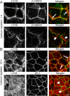
FIGURE 1:
FIGURE 1:
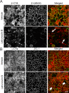
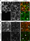
β-CYA is selectively enriched at newly formed epithelial junctions and the mature AJC. Confluent SK-CO15 cells were subjected to overnight extracellular calcium depletion to disassemble intercellular contacts, followed by either 1 (A) or 24 (B) h of calcium repletion to induce junctional reassembly. Cells were fixed and subjected to dual immunolabeling for either β-CYA or γ-CYA (red) with β-catenin and ZO-1 (green). Representative confocal images show accumulation of β-CYA at newly assembled junctions and the mature AJC (arrows) and lack of colocalization of junctional proteins with γ-CYA (arrowheads). Scale bar, 5 μm.
FIGURE 2:
FIGURE 2:
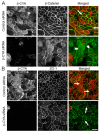
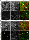
Apical β-CYA or γ-CYA structures have different sensitivity to latrunculin-induced F-actin depolymerization. Confluent SK-CO15 cell monolayers were incubated for 1 h with either vehicle or latrunculin B (1 μM). Cells were fixed and immunolabeled for cytoplasmic actins (red) and junctional protein (green). Note that latrunculin B treatment disrupted β-CYA labeling at apical junctions (arrows) but enhanced AJC localization of γ-CYA (arrowheads). Scale bar, 20 μm.
FIGURE 3:
FIGURE 3:
Down-regulation of β-CYA and γ-CYA attenuates formation of the paracellular barrier in apoptosis-independent manner. SK-CO15 cells were transfected with control, β-CYA–, or γ-CYA–specific siRNAs, and development of the paracellular barrier was examined by measuring TEER and fluoresceinated dextran flux. (A, B) Immunoblotting analysis shows a selective down-regulation of individual cytoplasmic actins on day 4 posttransfection, which differently affects the total actin level and does not stimulate apoptotic events such as PARP cleavage or caspases activation. Camptothecin treatment (10 μM for 24 h) is shown as a positive control for apoptosis. (C, D) Permeability assays reveal significant attenuation of TEER development and increased dextran fluxes in β-CYA– or γ-CYA–depleted cell monolayers on day 4 posttransfection. Data are presented as mean ± SE (n = 3); *p < 0.05, compared to control siRNA–transfected cells.
FIGURE 4:
FIGURE 4:
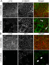
Down-regulation of β-CYA expression selectively disrupts the organization of mature AJs. SK-CO15 cells were transfected with either control or β-CYA–specific siRNAs, and the integrity of their AJs and TJs was examined by immunolabeling and confocal microscopy on day 4 posttransfection. Note that β-CYA depletion transforms junctional labeling of β-catenin (A, arrows) into a diffuse intracellular staining (arrowheads) but does not affect normal ZO-1 labeling at TJs (B, arrows). Scale bar, 20 μm.
FIGURE 5:
FIGURE 5:
Down-regulation of γ-CYA expression selectively impairs maintenance of TJs. SK-CO15 cells were transfected with either control or γ-CYA–specific siRNAs, and the integrity of their AJs and TJs was examined by immunolabeling and confocal microscopy on day 4 posttransfection. Note that depletion of γ-CYA disrupts ZO-1 labeling at TJs (arrowheads) without affecting β-catenin localization at mature AJs (arrows). Scale bar, 20 μm.
FIGURE 6:
FIGURE 6:
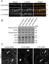
Down-regulation of β-CYA selectively impairs apicobasal cell polarity. (A) Reconstructed xz confocal images show proper localization of apical plasma membrane marker EBP50 (red) and basolateral membrane marker β-catenin (green) in control and γ-CYA–depleted SK-CO15 cells. By contrast, β-CYA–knockdown cells demonstrate diffuse intracellular staining of the apical plasma membrane marker. (B) Immunoblotting analysis shows that knockdown of β-CYA or γ-CYA did not affect expression of molecular constituents of the Par3–Par6–aPKC polarity complex, as well as phosphorylation of two aPKC isoforms. (C) Immunofluorescence images show junctional localization of Par-3 in control and γ-CYA–depleted cells (arrows) and mislocalization of Par-3 in β-CYA knockdown (arrowheads). Scale bar, 20 μm.
FIGURE 7:
FIGURE 7:
Down-regulation of β-CYA expression attenuates reassembly of AJs and TJs. Reassembly of AJs and TJs was examined in control and β-CYA–depleted SK-CO15 cells after 1 (A) or 3 (B) h of calcium repletion. Immunofluorescence labeling and confocal microscopy demonstrate accumulation of the majority of β-catenin and ZO-1 at the intercellular contacts in control cell monolayers (arrows), whereas in β-CYA–deficient cells a substantial amount of β-catenin remains in the cytoplasm and only short, defective ZO-1–based TJs are formed (arrowheads). Scale bar, 20 μm.
FIGURE 8:
FIGURE 8:
Down-regulation of γ-CYA expression attenuates reassembly of AJs and TJs. Junctional reassembly was examined in control and γ-CYA–depleted SK-CO15 cells after 1 (A) or 3 (B) h of calcium repletion. Immunofluorescence labeling and confocal microscopy show reassembly of the majority of β-catenin–based AJs and ZO-1–based TJs in control cell monolayers (arrows). By contrast, γ-CYA–deficient cells demonstrate only short, discontinuous AJs and TJs (arrowheads). Scale bar, 20 μm.
FIGURE 9:
FIGURE 9:
Down-regulation of β-CYA and γ-CYA attenuates assembly of the perijunctional actin cytoskeleton. Fluorescence labeling shows rapid formation of the circumferential F-actin belt at the level of apical junctions of control SK-CO15 cells after 3 h of calcium repletion (arrows). By contrast, β-CYA– (A) and γ-CYA–depleted (B) cells demonstrate decreased thickness and labeling intensity of perijunctional F-actin bundles (arrowheads). Scale bar, 20 μm.
FIGURE 10:
FIGURE 10:
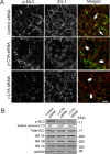
Down-regulation of β-CYA and γ-CYA inhibits activation of perijunctional NM II. (A) Immunofluorescence labeling shows significant accumulation of monophosphorylated myosin light chain (p-MLC) at newly assembled TJs in control SK-CO15 cells after 3 h of calcium repletion (arrows). By contrast, little accumulation of p-MLC at the areas of cell–cell contacts can be seen in either β-CYA– or γ-CYA–depleted cells (arrowheads). Scale bar, 20 μm. (B) Immunoblotting analysis shows the decreased cellular level of p-MLC in β-CYA– or γ-CYA–depleted SK-CO15 cells, whereas expression of total MLC, as well as NM II heavy chains A–C, remains unchanged. Data are presented as mean ± SE (n = 3); *p < 0.05, compared to control siRNA–transfected cells.
FIGURE 11:
FIGURE 11:
Knockdown of β-CYA and γ-CYA disrupts formation of intestinal epithelial cysts. (A) Immunoblotting analysis showing efficient knockdown of individual cytoplasmic actin isoform in Caco-2 cells. (B, C) Representative confocal images and quantitative analysis show formation of polarized cysts with well-defined lumen (arrow) in control siRNA–transfected Caco-2 cells embedded into 3D Matrigel. By contrast, formation of such hollow cysts was inhibited in Matrigel-growing β-CYA– or γ-CYA–depleted Caco-2 cells. Data are presented as mean ± SE (n = 3); *p < 0.05, compared to control siRNA–transfected cells. Scale bar, 5 μm.

References

    1. Abe K, Takeichi M. EPLIN mediates linkage of the cadherin catenin complex to F-actin and stabilizes the circumferential actin belt. Proc Natl Acad Sci USA. 2008;105:13–19. - PMC - PubMed
    1. Ammar DA, Nguyen PN, Forte JG. Functionally distinct pools of actin in secretory cells. Am J Physiol Cell Physiol. 2001;281:C407–C417. - PubMed
    1. Anderson JM, Van Itallie CM. Physiology and function of the tight junction. Cold Spring Harb Perspect Biol. 2009;1:a002584. - PMC - PubMed
    1. Ayscough K. Use of latrunculin-A, an actin monomer-binding drug. Methods Enzymol. 1998;298:18–25. - PubMed
    1. Belyantseva IA, et al. γ-Actin is required for cytoskeletal maintenance but not development. Proc Natl Acad Sci USA. 2009;106:9703–9708. - PMC - PubMed

Publication types

MeSH terms

LinkOut - more resources