. 2012 Jul 26:6:40.
doi: 10.3389/fnint.2012.00040. eCollection 2012.

The effects of cues on neurons in the basal ganglia in Parkinson's disease

Affiliations

The effects of cues on neurons in the basal ganglia in Parkinson's disease

Sridevi V Sarma et al. Front Integr Neurosci. .

Abstract

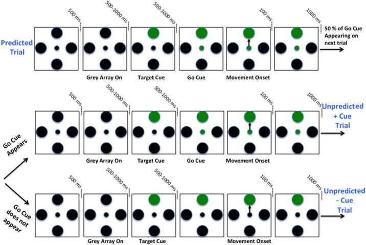
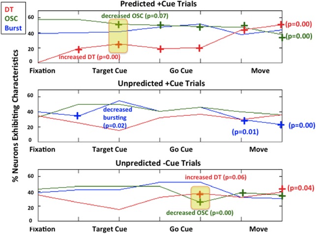
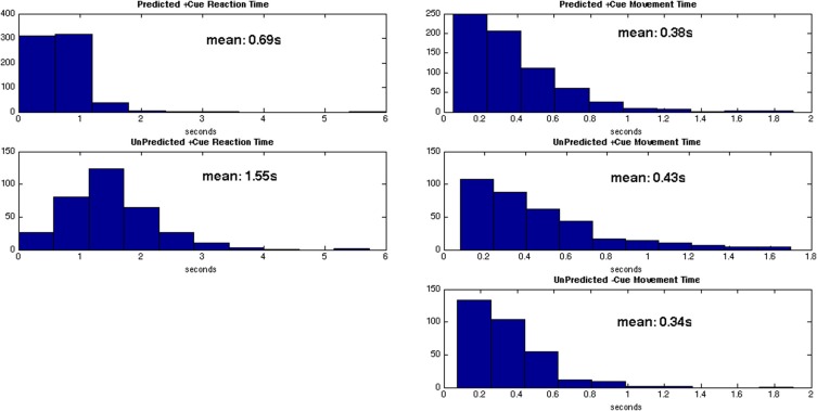
Visual cues open a unique window to the understanding of Parkinson's disease (PD). These cues can temporarily but dramatically improve PD motor symptoms. Although details are unclear, cues are believed to suppress pathological basal ganglia (BG) activity through activation of corticostriatal pathways. In this study, we investigated human BG neurophysiology under different cued conditions. We evaluated bursting, 10-30 Hz oscillations (OSCs), and directional tuning (DT) dynamics in the subthalamic nucleus (STN) activity while seven patients executed a two-step motor task. In the first step (predicted +cue), the patient moved to a target when prompted by a visual go cue that appeared 100% of the time. Here, the timing of the cue is predictable and the cue serves an external trigger to execute a motor plan. In the second step, the cue appeared randomly 50% of the time, and the patient had to move to the same target as in the first step. When it appeared (unpredicted +cue), the motor plan was to be triggered by the cue, but its timing was not predictable. When the cue failed to appear (unpredicted -cue), the motor plan was triggered by the absence of the visual cue. We found that during predicted +cue and unpredicted -cue trials, OSCs significantly decreased and DT significantly increased above baseline, though these modulations occurred an average of 640 ms later in unpredicted -cue trials. Movement and reaction times were comparable in these trials. During unpredicted +cue trials, OSCs, and DT failed to modulate though bursting significantly decreased after movement. Correspondingly, movement performance deteriorated. These findings suggest that during motor planning either a predictably timed external cue or an internally generated cue (generated by the absence of a cue) trigger the execution of a motor plan in premotor cortex, whose increased activation then suppresses pathological activity in STN through direct pathways, leading to motor facilitation in PD.

Keywords: Parkinson disease; cueing; neuromodulation; neuron; neuropathology.


Figures

Figure 1
Figure 1
Schematic of two-step sequential motor task. Predicted +Cue trial (Top), Unpredicted +Cue trial (middle), Unpredicted −Cue trial (bottom).
Figure 2
Figure 2
Time periods over which the CIF denoted by Equation (3), is estimated are shaded.
Figure 3
Figure 3
Optimal model parameters for an STN neuron during MV – and MV+ periods of a PD patient executing predicted-cued trials before movement (left) and after movement (right). Top row (movement direction modulation): optimal extrinsic factors eαd for d = 1, 2, 3, 4 (U,R,D,L) are plotted in black lines from left to right and corresponding 95% confidence intervals are shaded around each black line in a unique color for each direction. Middle row (short-term history modulation): optimal short-term history factors eβi for i = 1, 2,…, 10 are plotted in blue from right to left and the corresponding 95% confidence intervals are shaded in green. Bottom row (long-term history modulation) optimal long-term history factors eγj for j = 1, 2,…, 14 are plotted in blue from right to left and corresponding 95% confidence intervals are shaded in green. Note the change in time scale for bottom row.
Figure 4
Figure 4
Raw spike train data from a single STN neuron in PD patient. The spike train is in blue and the corresponding estimate of the CIF is in red.
Figure 5
Figure 5
Modulations of each characteristic for each trial type. Predicted +Cue Trials (top); Unpredicted +Cue Trials (middle); Unpredicted −Cue Trials (bottom). When the percentage of neurons exhibit neuronal spiking characteristics in a monotonically decreasing less pathological direction (decreasing beta oscillations, increasing directional tuning) for the duration of the trial, we denote that with a “+” symbol.
Figure 6
Figure 6
Distributions for reaction times (left) and movement times (right) for each trial type.

References

    1. Abosch A., Hutchison W. D., Saint-Cyr J. A., Dostrovsky J. O., Lozano A. M. (2002). Movement related neurons of the subthalamic nucleus in patients with Parkinson disease. J. Neurosurg. 97, 1167–1172 10.3171/jns.2002.97.5.1167 - DOI - PubMed
    1. Alexander G. E., Crutcher M. D. (1990). Preparation for movement: neural representations of intended direction in three motor areas of the monkey. J. Neurophysiol. 64, 133–150 - PubMed
    1. Amirnovin R., Williams Z. M., Cosgrove G. R., Eskandar E. N. (2004). Visually guided movements suppress subthalamic oscillations in Parkinson's disease patients. J. Neurosci. 24, 11302–11306 10.1523/JNEUROSCI.3242-04.2004 - DOI - PMC - PubMed
    1. Amirnovin R., Williams Z. M., Cosgrove G. R., Eskandar E. N. (2006). Experience with microelectrode guided subthalamic nucleus deep brain stimulation. Neurosurgery 58(Suppl. 1), ONS96–ONS102 10.1227/01.NEU.0000192690.45680.C2 - DOI - PubMed
    1. Azulay J. P., Mesure S., Amblard B., Blin O., Sangla I., Pouget J. (1999). Visual control of locomotion in Parkinson's disease. Brain 122, 111–120 10.1093/brain/122.1.111 - DOI - PubMed

LinkOut - more resources