. 2012 Apr;5(2):103-109.
doi: 10.7156/v5i2p103.

Pathology Imaging Informatics for Clinical Practice and Investigative and Translational Research

Affiliations

Pathology Imaging Informatics for Clinical Practice and Investigative and Translational Research

Evita T Sadimin et al. N Am J Med Sci (Boston). 2012 Apr.

Abstract

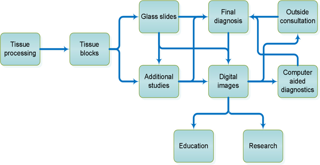
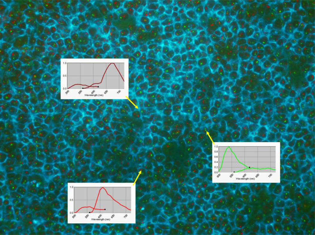
Pathologists routinely interpret gross and microscopic specimens to render diagnoses and to engage in a broad spectrum of investigative research. Multiple studies have demonstrated that imaging technologies have progressed to a level at which properly digitized specimens provide sufficient quality comparable to the traditional glass slides examinations. Continued advancements in this area will have a profound impact on the manner in which pathology is conducted from this point on. Several leading institutions have already undertaken ambitious projects directed toward digitally imaging, archiving, and sharing pathology specimens. As a result of these advances, the use of informatics in diagnostic and investigative pathology applications is expanding rapidly. In addition, the advent of novel technologies such as multispectral imaging makes it possible to visualize and analyze imaged specimens using multiple wavelengths simultaneously. As these powerful technologies become increasingly accepted and adopted, the opportunities for gaining new insight into the underlying mechanisms of diseases as well as the potential for discriminating among subtypes of pathologies are growing accordingly.


Conflict of interest statement

CONFLICT OF INTEREST

None.

Figures

Figure 1
Figure 1
Anatomical pathology workflow incorporating imaging informatics into clinical practice, investigative research and education.
Figure 2
Figure 2
Combined CD20 immunofluorescence (membrane staining) and (11;14) FISH of a germinal center in a frozen tonsil. The wavelength of each component detected by the multispectral camera is shown as follows: CD20 (top inset) - emission: 680nm, green FISH (middle inset) - emission: 540nm, and red FISH (bottom inset) - emission: 610nm. The emission for CD20 is near infrared and is not readily visible to human eyes but is easily detected by the camera. To provide the best color contrast, the CD20 shown on the image has been pseudo-colored cyan. (The figure is available at http://pleiad.umdnj.edu/CBII/images/NAJMS_image_2.png in higher resolution.)

References

    1. Hedvat CV. Digital microscopy: past, present, and future. Arch Pathol Lab Med. 2010;134(11):1666–1670. - PubMed
    1. Pinco J, Goulart RA, Otis CN, Garb J, Pantanowitz L. Impact of digital image manipulation in cytology. Arch Pathol Lab Med. 2009;133(1):57–61. - PubMed
    1. Yagi Y, Gilbertson JR. The importance of optical optimization in whole slide imaging (WSI) and digital pathology imaging. Diagnostic pathology. 2008;3(Suppl I):S1. - PMC - PubMed
    1. Martina JD, Simmons C, Jukic DM. High-definition hematoxylin and eosin staining in a transition to digital pathology. J Pathol Inform. 2011;2:45. - PMC - PubMed
    1. Thrall M, Pantanowitz L, Khalbuss W. Telecytology: Clinical applications, current challenges, and future benefits. J Pathol Inform. 2011;2:51. - PMC - PubMed

LinkOut - more resources