. 2012 Sep 1;189(5):2584-96.
doi: 10.4049/jimmunol.1200999. Epub 2012 Aug 1.

Dendritic cell sphingosine 1-phosphate receptor-3 regulates Th1-Th2 polarity in kidney ischemia-reperfusion injury

Affiliations

Dendritic cell sphingosine 1-phosphate receptor-3 regulates Th1-Th2 polarity in kidney ischemia-reperfusion injury

Amandeep Bajwa et al. J Immunol. .

Abstract

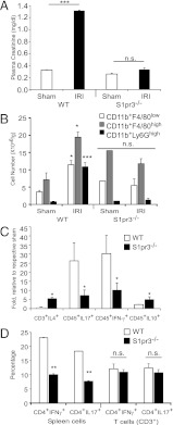
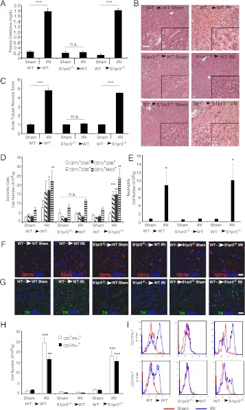
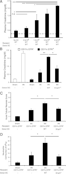
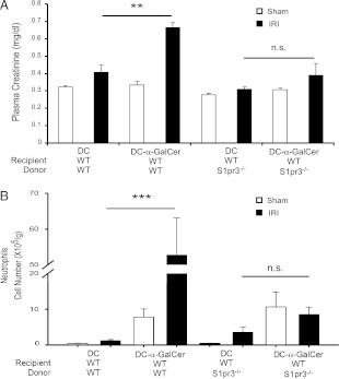
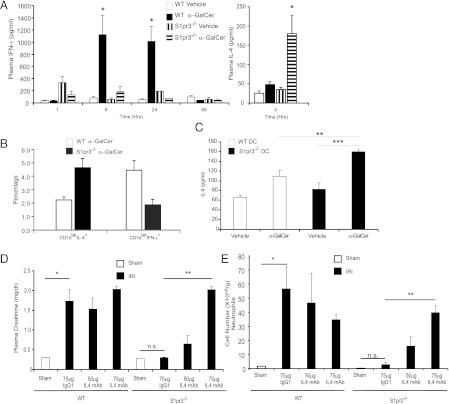
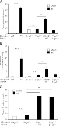
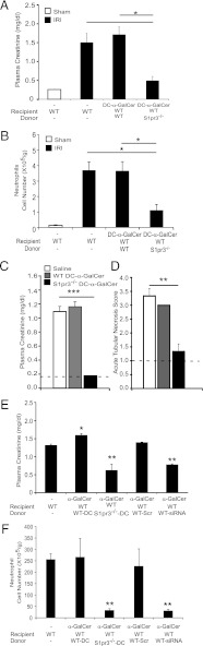
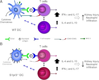
Dendritic cells (DCs) are central to innate and adaptive immunity of early kidney ischemia-reperfusion injury (IRI), and strategies to alter DC function may provide new therapeutic opportunities. Sphingosine 1-phosphate (S1P) modulates immunity through binding to its receptors (S1P1-5), and protection from kidney IRI occurs in S1P3-deficient mice. Through a series of experiments we determined that this protective effect was owing in part to differences between S1P3-sufficient and -deficient DCs. Mice lacking S1P3 on bone marrow cells were protected from IRI, and S1P3-deficient DCs displayed an immature phenotype. Wild-type (WT) but not S1P3-deficient DCs injected into mice depleted of DCs prior to kidney IR reconstituted injury. Adoptive transfer (i.e., i.v. injection) of glycolipid (Ag)-loaded WT but not S1P3-deficient DCs into WT mice exacerbated IRI, suggesting that WT but not S1P3-deficient DCs activated NKT cells. Whereas WT DC transfers activated the Th1/IFN-γ pathway, S1P3-deficient DCs activated the Th2/IL-4 pathway, and an IL-4-blocking Ab reversed protection from IRI, supporting the concept that IL-4 mediates the protective effect of S1P3-deficient DCs. Administration of S1P3-deficient DCs 7 d prior to or 3 h after IRI protected mice from IRI and suggests their potential use in cell-based therapy. We conclude that absence of DC S1P3 prevents DC maturation and promotes a Th2/IL-4 response. These findings highlight the importance of DC S1P3 in modulating NKT cell function and IRI and support development of selective S1P3 antagonists for tolerizing DCs for cell-based therapy or for systemic administration for the prevention and treatment of IRI and autoimmune diseases.


Figures

FIGURE 1.
FIGURE 1.
S1pr3−/− mice are protected from kidney IRI. Kidney IRI (26 min ischemia and 24 h reperfusion) was performed in WT and S1pr3−/− mice. (A) There was no difference in baseline plasma creatinine between sham-operated WT and S1pr3−/− mice. WT mice have a significant rise in plasma creatinine compared with S1pr3−/− mice after IRI (n = 4–6/group). ***p < 0.001. (B) FACS analysis of total live (7-AAD) leukocytes (CD45+) gated for CD11b+F4/80low (macrophages), CD11b+F4/80high (DCs), and CD11b+Ly6Ghigh (neutrophils) in WT and S1pr3−/− mice after IRI (n = 4–6/group). ***p < 0.001 and *p < 0.05 compared with respective sham-operated mice. (C) FACS analysis of total live (7-AAD) leukocytes (CD45+) and T cells (CD3+) expressing pro- (IL-17, IFN-γ) and anti-inflammatory (IL-4, IL-10) cytokines after IRI (n = 2–5/group). *p < 0.05 compared with WT mice. (D) Leukocyte expression of IL-17 and IFN-γ in WT and S1pr3−/− total spleen cells and T cell subset (CD3+) cultured under Th17-inducing conditions. Data are expressed as percentage of total CD4+ cells (n = 3–5/group). **p < 0.01 compared with WT mice. Data are expressed as means ± SEM.
FIGURE 2.
FIGURE 2.
Kidneys of mice with S1P3-deficient BM-derived cells (S1pr3−/−→WT) are protected from IRI. Kidney IRI (28 min ischemia, 24 h reperfusion) was performed in BM chimeric mice: WT→WT, S1pr3−/−→WT and WT→ S1pr3−/−. (A) Plasma creatinine levels (n = 8–10/group). ***p < 0.001. (B) H&E staining of kidney sections after IRI; insets show a ×2.5 magnified image. Scale bar, 100 μm. (C) Semiquantitative measure of tubular injury in H&E-stained kidney sections using a scale of 0–5 as described in Materials and Methods (n = 8–10/group). ***p < 0.001. (D) FACS analysis of DC subsets of total live (7-AAD) leukocytes (CD45+) (n = 6–8/group). *p < 0.05, **p < 0.01, and ***p < 0.001 versus respective sham-operated mice. (E) FACS analysis of total live neutrophils (CD11b+GR-1high) (n = 6–8/group). *p < 0.05 versus respective sham-operated mice. (F and G) Immunofluorescence labeling of DCs (CD11c; red) (F) in the kidney outer medulla or neutrophils (7/4, green) or (G) in kidney sections of chimeric mice after sham or IRI; nuclei are labeled with DAPI (blue). Scale bars, 40 μm. (H) FACS analysis of T cell (CD3+) and non-T cell (CD3) subsets of total live IFN-γ–producing leukocytes in kidney after IRI (n = 6–8/group). **p < 0.01 and ***p < 0.001 versus respective sham-operated mice. (I) Representative histograms (total live leukocytes gated on CD3+ and CD3 cells that produce IFN-γ) show a rightward shift after IR (red) compared with sham (blue), indicative of increased IFN-γ–producing cells in WT→WT and WT→S1pr3−/− mice but not in S1pr3−/−→WT mice. Data are expressed as means ± SEM.
FIGURE 3.
FIGURE 3.
Kidney injury after IR is blunted in DC-depleted mice reconstituted with S1pr3−/− BMDCs. (A) Injection (i.v.) of increasing numbers of WT BMDCs to WT mice 18 h prior to IRI (24 min ischemia) induces kidney injury (n = 4–6/group). *p < 0.05, ***p < 0.001. (BD) DCs were depleted in DT-sensitive, human DTR-expressing mice (CD11c-DTR+) but not in CD11c-DTR mice by treatment with DT; 20 h later mice were injected with either 1× PBS (−), DCs from WT mice, or DCs from S1pr3−/− mice, and mice were subjected to IR (26 min ischemia) 6 h after DC injection. (B) CD11c-DTR+ mice reconstituted with WT DCs had more injury after IR than did CD11c-DTR+ mice with no DCs or with S1pr3−/− DCs. (C) Semiquantitative measure of tubular injury in H&E-stained kidney sections. (D) Reconstitution of DC-depleted CD11c-DTR+ mice with WT but not S1pr3−/− DCs increased neutrophil infiltration in kidneys (n = 3–6/group). Data are expressed as means ± SEM. *p < 0.05, **p < 0.01.
FIGURE 4.
FIGURE 4.
S1pr3−/− BMDCs loaded with α-GalCer are unable to induce injury after IR. (A) Mild ischemia (24 min) did not increase plasma creatinine. Injection of α-GalCer–loaded WT but not S1pr3−/− DCs caused a significant increase in plasma creatinine (A) and neutrophil infiltration (B) after mild IR (n = 4–9/group). Data are expressed as means ± SEM. **p < 0.01, ***p < 0.001.
FIGURE 5.
FIGURE 5.
Protection of S1pr3−/− mice from IRI is IL-4–dependent. (A) Plasma levels of IFN-γ 1–48 h and IL-4 4 h after treatment of WT and S1pr3−/− mice with vehicle or α-GalCer (10 μg/mouse, i.p.) (n = 3–4/group). *p < 0.05 compared with respective vehicle control for each time point. (B) WT and S1pr3−/− mice were injected with α-GalCer (1 μg/mouse, i.v.), and liver NKT cell (CD1dtet) IFN-γ and IL-4 levels were measured by FACS. Compared to WT mice, S1pr3−/− mice had a higher percentage of liver CD1dtet-positive cells that made IL-4, and WT mice had higher percentage of CD1dtet-positive cells that made IFN-γ compared with S1pr3−/− mice. (C) DCs from WT and S1pr3−/− mice were loaded with vehicle or α-GalCer prior to coculture with NKT1.2 hybridoma cells, and IL-4 was measured in the culture medium. Little or no measurable IL-2 or IL-4 was detected in culture medium of WT DCs or NKT1.2 cells that were cultured alone (n = 3/group). **p < 0.01, ***p < 0.001. (D and E) WT or S1pr3−/− mice were injected with neutralizing IL-4 mAb (50, 75, or 100 μg) or isotype control Ab (75 μg) 1 d prior to kidney ischemia (26 min), and plasma creatinine (D) and kidney neutrophil infiltration (by flow cytometry) (E) were measured after 20 h of reperfusion (n = 3/group). Data are expressed as means ± SEM. *p < 0.05, **p < 0.01.
FIGURE 6.
FIGURE 6.
Injection of WT DCs in S1pr3−/− mice reconstitutes injury after IR. (A) Plasma creatinine increased after IRI (26 min ischemia) in WT but not S1pr3−/− mice treated with vehicle. Injury was partially restored in S1pr3−/− mice after injection of WT but not S1pr3−/− DCs (0.5 × 10⁶ cells were injected 24 h prior to surgery) as demonstrated by plasma creatinine (A) and neutrophil infiltration (B) (n = 3–4/group). *p < 0.05, **p < 0.01, ***p < 0.001. (C) Reconstitution of Rag-1−/− mice with WT or S1pr3−/− T conventional (CD4+CD25) cells results in significantly higher plasma creatinine after kidney IRI (n = 3–4/group). Data are expressed as means ± SEM. **p < 0.01.
FIGURE 7.
FIGURE 7.
S1pr3−/− DCs are effective both in preventing kidney IRI and in treating injury after IR. (A and B) WT mice were injected (i.v.) with WT DCs or S1pr3−/− DCs (0.5 × 10⁶; loaded with α-GalCer) 7 d prior to IRI (26 min). (A) Plasma creatinine; (B) neutrophil infiltration in kidneys (n = 3–4/group). *p < 0.05, **p < 0.01, ***p < 0.001. (C) WT mice were subjected to 26 min ischemia and 24 h reperfusion and injected (i.v.) with WT DCs or S1pr3−/− DCs (0.5 × 10⁶; loaded with α-GalCer) 3 h after ischemia; plasma creatinine was measured at the 24 h reperfusion. Dashed line, creatinine levels in sham-operated mice (n = 3–4). p < 0.001. (D) Semiquantitative measure of tubular injury in H&E-stained kidney sections. Dashed line, ATN in sham-operated mice. (E) Knockdown of S1pr3 in WT DCs results in less kidney injury in WT mice as indicated by plasma creatinine and (F) neutrophil infiltration. WT DCs transfected with S1P3 siRNA (WT-siRNA) or scrambled oligonucleotides (WT-Scr) or untreated WT or S1pr3−/− DCs were loaded with α-GalCer and injected into WT mice (0.5 × 10⁶ cells) 24 h prior to IRI (n = 3–4/group). Data are means ± SEM. *p < 0.05, **p < 0.01.
FIGURE 8.
FIGURE 8.
Absence of S1P3 in DCs polarizes T cells to a Th2 phenotype in kidney IRI. In kidney IRI, (A) WT DCs via their class I-like CD1d molecule present endogenous glycolipid or α-GalCer to NKT cells and along with CD40 costimulatory molecule–CD40L interaction cause– NKT cell activation. Additionally, DCs can interact with and activate conventional T cells and regulatory T cells through a variety of mechanisms. Activated NKT cells produce large amounts of IFN-γ (Th1 response), leading to neutrophil infiltration and kidney injury. (B) DCs lacking S1P3 (S1pr3−/− DCs) also present endogenous glycolipid or α-GalCer to NKT cells via their class I-like CD1d molecule but have reduced CD40 and cytokine/chemokine expression after kidney IRI. NKT cells stimulated by α-GalCer–loaded S1pr3−/− DCs produce large amounts of IL-4 (Th2 response) and IL-10 with low to minimal IFN-γ (Th1) and IL-17. Similarly, S1pr3−/− DCs in mice subject to kidney IRI may also fail to induce a Th1 response in conventional T cells and hence promote increased IL-4 production by conventional T cells. High levels of IL-4 result in less neutrophil infiltration and less kidney injury. Neutralization of IL-4 with blocking mAb reverses this protective effect of IL-4 in S1pr3−/− mice, leading to more kidney injury. The present studies focused on DC/NKT interactions in IRI, but other mechanisms, such as reduced IL-17 or increased IL-10, may also contribute to protection in S1pr3−/− mice and in WT mice treated with S1pr3−/− DCs prior to IRI. Mechanisms underlying the beneficial effects of S1pr3−/− DCs administered after established IRI have not yet been identified and could include enhanced repair processes.

References

    1. Bonventre J. V., Yang L. 2011. Cellular pathophysiology of ischemic acute kidney injury. J. Clin. Invest. 121: 4210–4221 - PMC - PubMed
    1. Grenz A., Bauerle J. D., Dalton J. H., Ridyard D., Badulak A., Tak E., McNamee E. N., Clambey E., Moldovan R., Reyes G., et al. 2012. Equilibrative nucleoside transporter 1 (ENT1) regulates postischemic blood flow during acute kidney injury in mice. J. Clin. Invest. 122: 693–710 - PMC - PubMed
    1. Li L., Huang L., Sung S. S., Vergis A. L., Rosin D. L., Rose C. E., Jr., Lobo P. I., Okusa M. D. 2008. The chemokine receptors CCR2 and CX3CR1 mediate monocyte/macrophage trafficking in kidney ischemia-reperfusion injury. Kidney Int. 74: 1526–1537 - PMC - PubMed
    1. Nelson P. J. 2007. Renal ischemia-reperfusion injury: renal dendritic cells loudly sound the alarm. Kidney Int. 71: 604–605 - PubMed
    1. Soos T. J., Sims T. N., Barisoni L., Lin K., Littman D. R., Dustin M. L., Nelson P. J. 2006. CX3CR1+ interstitial dendritic cells form a contiguous network throughout the entire kidney. Kidney Int. 70: 591–596 - PubMed

Publication types

MeSH terms

Substances