

Real-time MRI-guided right heart catheterization in adults using passive catheters
- PMID: 22855740
- PMCID: PMC3561614
- DOI: 10.1093/eurheartj/ehs189
Real-time MRI-guided right heart catheterization in adults using passive catheters
Abstract
Aims: Real-time MRI creates images with superb tissue contrast that may enable radiation-free catheterization. Simple procedures are the first step towards novel interventional procedures. We aim to perform comprehensive transfemoral diagnostic right heart catheterization in an unselected cohort of patients entirely using MRI guidance.
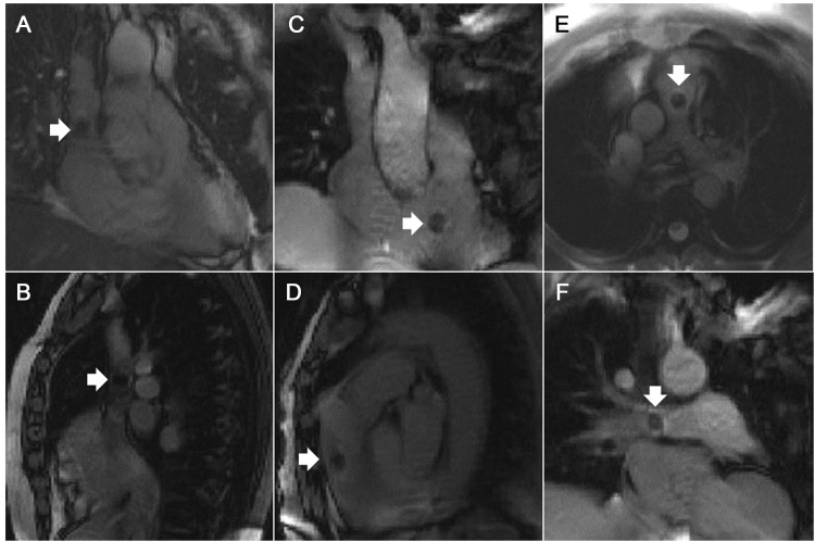
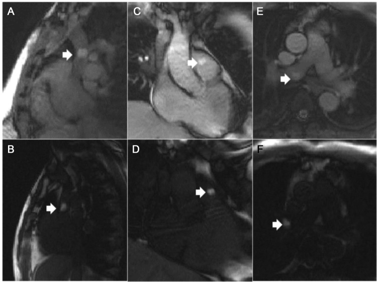
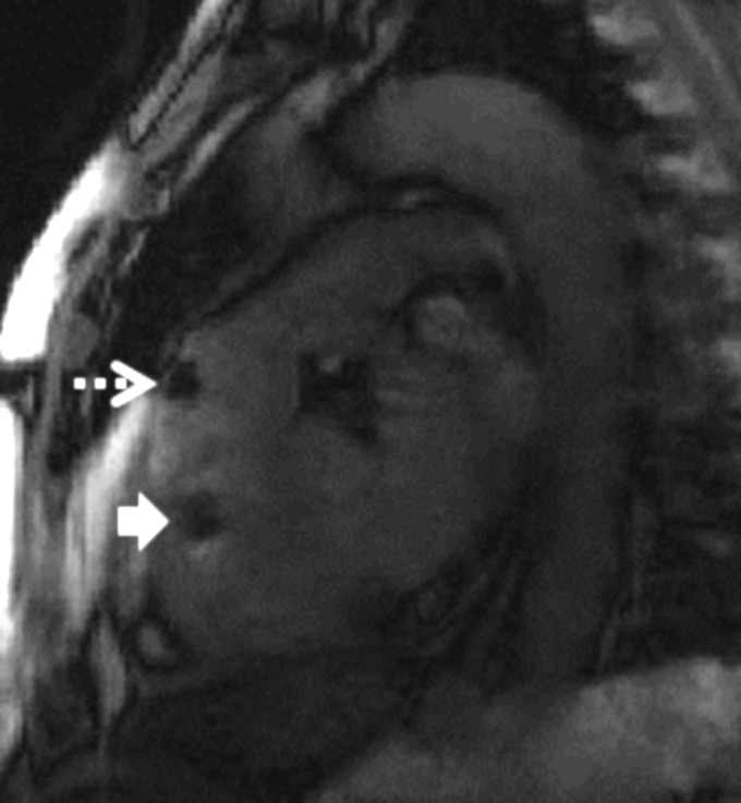
Methods and results: We performed X-ray and MRI-guided transfemoral right heart catheterization in consecutive patients undergoing clinical cardiac catheterization. We sampled both cavae and both pulmonary arteries. We compared success rate, time to perform key steps, and catheter visibility among X-ray and MRI procedures using air-filled or gadolinium-filled balloon-tipped catheters. Sixteen subjects (four with shunt, nine with coronary artery disease, three with other) underwent paired X-ray and MRI catheterization. Complete guidewire-free catheterization was possible in 15 of 16 under both. MRI using gadolinium-filled balloons was at least as successful as X-ray in all procedure steps, more successful than MRI using air-filled balloons, and better than both in entering the left pulmonary artery. Total catheterization time and individual procedure steps required approximately the same amount of time irrespective of image guidance modality. Catheter conspicuity was best under X-ray and next-best using gadolinium-filled MRI balloons.
Conclusion: In this early experience, comprehensive transfemoral right heart catheterization appears feasible using only MRI for imaging guidance. Gadolinium-filled balloon catheters were more conspicuous than air-filled ones. Further workflow and device enhancement are necessary for clinical adoption.
Figures

Comment in
-
Interventional vascular MRI: moving forward.Eur Heart J. 2013 Feb;34(5):327-9. doi: 10.1093/eurheartj/ehs236. Epub 2012 Nov 26. Eur Heart J. 2013. PMID: 23184892 No abstract available.
References
-
- Sorensen TS, Atkinson D, Schaeffter T, Hansen MS. Real-time reconstruction of sensitivity encoded radial magnetic resonance imaging using a graphics processing unit. IEEE Trans Med Imaging. 2009;28:1974–1985. - PubMed
-
- Breuer FA, Kellman P, Griswold MA, Jakob PM. Dynamic autocalibrated parallel imaging using temporal GRAPPA (TGRAPPA) Magn Reson Med. 2005;53:981–985. - PubMed
-
- Razavi R, Hill DL, Keevil SF, Miquel ME, Muthurangu V, Hegde S, Rhode K, Barnett M, van Vaals J, Hawkes DJ, Baker E. Cardiac catheterisation guided by MRI in children and adults with congenital heart disease. Lancet. 2003;362:1877–1882. - PubMed
Publication types
MeSH terms
Substances
Grants and funding
LinkOut - more resources
Full Text Sources
Other Literature Sources
Medical
