. 2012 Oct;108(8):2173-8.
doi: 10.1152/jn.00505.2012. Epub 2012 Aug 1.

Transcranial electrical stimulation over visual cortex evokes phosphenes with a retinal origin

Affiliations

Transcranial electrical stimulation over visual cortex evokes phosphenes with a retinal origin

Kohitij Kar et al. J Neurophysiol. 2012 Oct.

Abstract

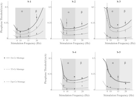
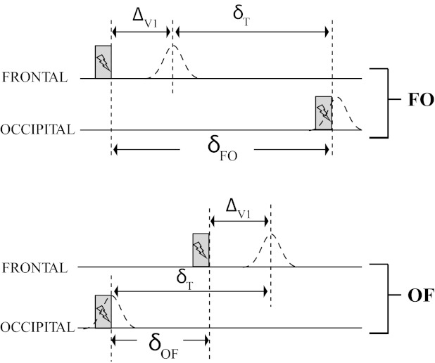
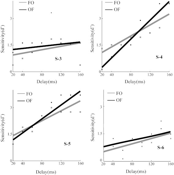
Transcranial electrical stimulation (tES) is a promising therapeutic tool for a range of neurological diseases. Understanding how the small currents used in tES spread across the scalp and penetrate the brain will be important for the rational design of tES therapies. Alternating currents applied transcranially above visual cortex induce the perception of flashes of light (phosphenes). This makes the visual system a useful model to study tES. One hypothesis is that tES generates phosphenes by direct stimulation of the cortex underneath the transcranial electrode. Here, we provide evidence for the alternative hypothesis that phosphenes are generated in the retina by current spread from the occipital electrode. Building on the existing literature, we first confirm that phosphenes are induced at lower currents when electrodes are placed farther away from visual cortex and closer to the eye. Second, we explain the temporal frequency tuning of phosphenes based on the well-known response properties of primate retinal ganglion cells. Third, we show that there is no difference in the time it takes to evoke phosphenes in the retina or by stimulation above visual cortex. Together, these findings suggest that phosphenes induced by tES over visual cortex originate in the retina. From this, we infer that tES currents spread well beyond the area of stimulation and are unlikely to lead to focal neural activation. Novel stimulation protocols that optimize current distributions are needed to overcome these limitations of tES.


Figures

Fig. 1.
Fig. 1.
Design of the phosphene detection task. Trials consisted of 2 intervals (1 and 2) separated by a beep; transcranial alternating current stimulation (tACS) was applied in 1 of the 2 intervals. Subjects reported the interval in which they saw phosphenes, and we determined the current level at which the subject's phosphene detection performance reached 75% correct. “STIM” refers to the time of application of transcranial electrical stimulation as indicated by the lightning bolt.
Fig. 2.
Fig. 2.
Threshold measurement and frequency tuning. Dependence of phosphene thresholds on stimulation frequency (8–20 Hz) and electrode position for 5 subjects (S-1, S-2, S-3, S-4, and S-5). The α- and β-frequency bands are shaded in dark and light gray, respectively. Consistent with the findings of Kanai et al. (2008), the optimal stimulation frequency for these experiments done in near darkness was typically in the α-range. The box and “x” markers are the estimated thresholds; error bars indicate 95% confidence intervals, and the lines represent the model fit based on the retinal ganglion cell model. The figure shows that current thresholds decreased as the electrode was moved closer to the eye, consistent with a retinal origin. Moreover, it shows that the temporal frequency tuning is consistent with the known properties of primate retinal ganglion cells.
Fig. 3.
Fig. 3.
Predictions for the double-pulse detection task based on the hypothesis that occipital stimulation evokes neural activity directly in the underlying cortex. Gray bars represent the stimulation, and dashed curves the activity in V1 under the cortical hypothesis. This figure illustrates why the cortical hypothesis predicts large differences in the time needed to distinguish 2 phosphenes between Oz-Fpz (OF) and Fpz-Oz (FO) stimulation. ΔV1, the time it takes for a stimuli to reach the visual cortex from the retina; δT, the minimum time to perceive 2 consecutive phosphenes. See main text for a full description.
Fig. 4.
Fig. 4.
Double-pulse detection sensitivity. Behavioral report for 4 subjects (S-3, S-4, S-5, and S-6) in the double-pulse detection task. The sensitivity (d′) of the subject to detect 2 flashes is shown as a function of the time between the 2 stimulation pulses. The bold lines are lines fitted to the data points. Threshold delay is defined as the delay at which d′ reached 1.5. The threshold delays for OF and FO conditions were statistically indistinguishable for all subjects (permutation test, P > 0.05).

References

    1. Attwell D. Interaction of low frequency electric fields with the nervous system: the retina as a model system. Radiat Prot Dosimetry 106: 341–348, 2003 - PubMed
    1. Baylor DA, Nunn BJ, Schnapf JL. The photocurrent, noise and spectral sensitivity of rods of the monkey Macaca fascicularis. J Physiol 357: 575–607, 1984 - PMC - PubMed
    1. Benardete EA, Kaplan E. The dynamics of primate M retinal ganglion cells. Vis Neurosci 16: 355–368, 1999 - PubMed
    1. Brindley GS. The site of electrical excitation of the human eye. J Physiol 127: 189–200, 1955 - PMC - PubMed
    1. Datta A, Bansal V, Diaz J, Patel J, Reato D, Bikson M. Gyri-precise head model of transcranial direct current stimulation: improved spatial focality using a ring electrode versus conventional rectangular pad. Brain Stimul 2: 201–207, 207.e1, 2009 - PMC - PubMed

Publication types

LinkOut - more resources