. 2012 Aug 1;32(31):10479-83.
doi: 10.1523/JNEUROSCI.0803-12.2012.

The resting transducer current drives spontaneous activity in prehearing mammalian cochlear inner hair cells

Affiliations

The resting transducer current drives spontaneous activity in prehearing mammalian cochlear inner hair cells

Stuart L Johnson et al. J Neurosci. .

Abstract

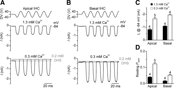
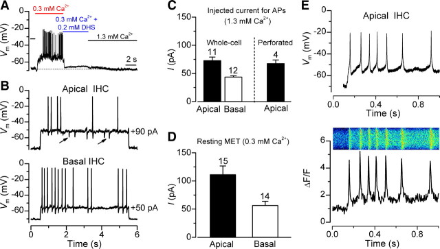
Spontaneous Ca(2+)-dependent electrical activity in the immature mammalian cochlea is thought to instruct the formation of the tonotopic map during the differentiation of sensory hair cells and the auditory pathway. This activity occurs in inner hair cells (IHCs) during the first postnatal week, and the pattern differs along the cochlea. During the second postnatal week, which is before the onset of hearing in most rodents, the resting membrane potential for IHCs is apparently more hyperpolarized (approximately -75 mV), and it remains unclear whether spontaneous action potentials continue to occur. We found that when mouse IHC hair bundles were exposed to the estimated in vivo endolymphatic Ca(2+) concentration (0.3 mm) present in the immature cochlea, the increased open probability of the mechanotransducer channels caused the cells to depolarize to around the action potential threshold (approximately -55 mV). We propose that, in vivo, spontaneous Ca(2+) action potentials are intrinsically generated by IHCs up to the onset of hearing and that they are likely to influence the final sensory-independent refinement of the developing cochlea.


Figures

Figure 1.
Figure 1.
Mechanoelectrical transducer current in cochlear IHCs. A, B, Saturating mechanotransducer currents recorded from an apical and basal mouse IHC, respectively, exposed to 1.3 mm Ca2+ (middle) and an endolymphatic Ca2+ concentration (0.3 mm: bottom). The driver voltage (DV) signal to the fluid jet is shown above the traces (positive deflection of the DV is inhibitory). Note that mechanotransducer current amplitude was larger and its fraction activated at rest was increased in the presence of endolymph-like Ca2+. In 0.3 mm Ca2+ the mechanotransducer current was abolished by 0.2 mm DHS. IHC holding potential was −84 mV. C, D, Maximum amplitude (C) and resting open probability (D) of the mechanotransducer current in perilymph-like (1.3 mm) and endolymph-like (0.3 mm: see Materials and Methods for details) Ca2+ concentrations. The resting open probability was calculated by dividing the mechanotransducer current available at rest (the difference between the current level before the stimulus, indicated by the dashed line, and the current level on the negative phase of the stimulus when all channels were closed) by the maximum peak-to-peak mechanotransducer current.
Figure 2.
Figure 2.
Action potential activity in second-postnatal week IHCs. A, IHC voltage responses recorded during the superfusion of 1.3 mm Ca2+ or 0.3 mm Ca2+ alone or together with DHS. B, Action potentials elicited from an apical (top) and a basal (bottom) IHC during depolarizing current injection in 1.3 mm Ca2+. Arrows indicate IPSPs. C, Average current amplitude required to depolarize apical (whole-cell and perforated patch recordings) and basal (whole-cell recordings) IHCs to trigger action potential activity in 1.3 mm Ca2+. D, Average resting mechanotransducer current in apical and basal IHCs in 0.3 mm Ca2+. This was obtained at −70 mV, which matches the IHC resting membrane potential in 1.3 mm Ca2+. E, Simultaneous recording of induced action potential activity (top) and calcium transients (bottom) in an apical IHC. Minimum current injection was used to reach spike threshold. The Ca2+ image was taken across the base of the IHC and has been smoothed in ImageJ.

References

    1. Beutner D, Moser T. The presynaptic function of mouse cochlear inner hair cells during development of hearing. J Neurosci. 2001;21:4593–4599. - PMC - PubMed
    1. Blankenship AG, Feller MB. Mechanisms underlying spontaneous patterned activity in developing neural circuits. Nat Rev Neurosci. 2010;11:18–29. - PMC - PubMed
    1. Bosher SK, Warren RL. A study of the electrochemistry and osmotic relationships of the cochlear fluids in the neonatal rat at the time of the development of the endocochlear potential. J Physiol. 1971;212:739–761. - PMC - PubMed
    1. Bosher SK, Warren RL. Very low calcium content of cochlear endolymph, an extracellular fluid. Nature. 1978;273:377–378. - PubMed
    1. Heidrych P, Zimmermann U, Kuhn S, Franz C, Engel J, Duncker SV, Hirt B, Pusch CM, Ruth P, Pfister M, Marcotti W, Blin N, Knipper M. Otoferlin interacts with myosin VI: implications for maintenance of the basolateral synaptic structure of the inner hair cell. Hum Mol Genet. 2009;18:2779–2790. - PubMed

Publication types

MeSH terms

Substances

LinkOut - more resources