

Deconstructing the architecture of dorsal and ventral attention systems with dynamic causal modeling
- PMID: 22855813
- PMCID: PMC3432566
- DOI: 10.1523/JNEUROSCI.0414-12.2012
Deconstructing the architecture of dorsal and ventral attention systems with dynamic causal modeling
Abstract
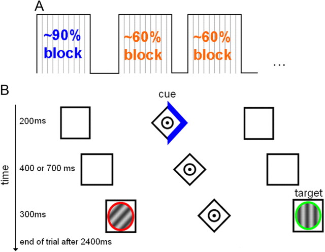
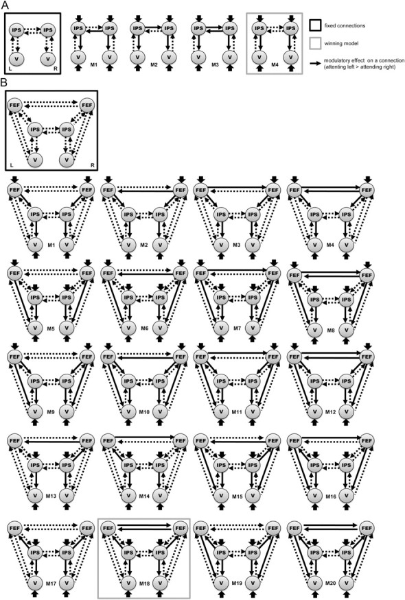
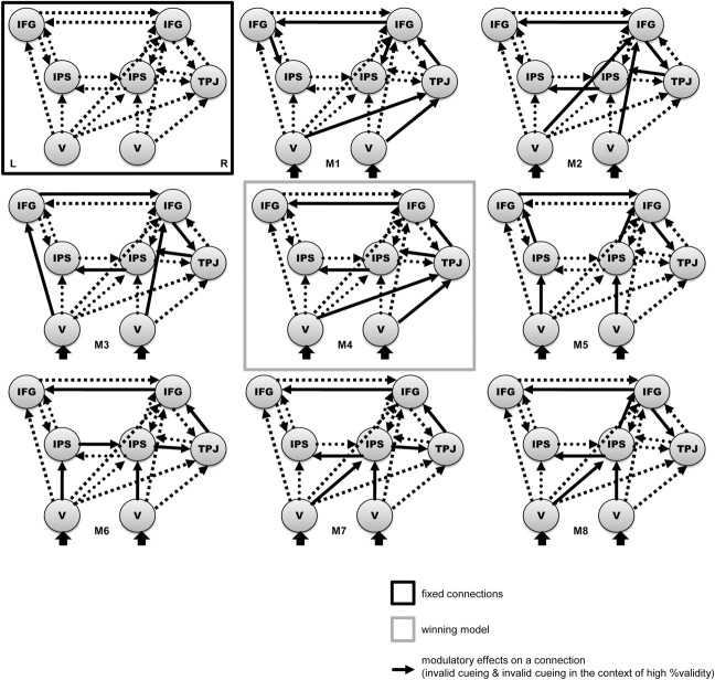
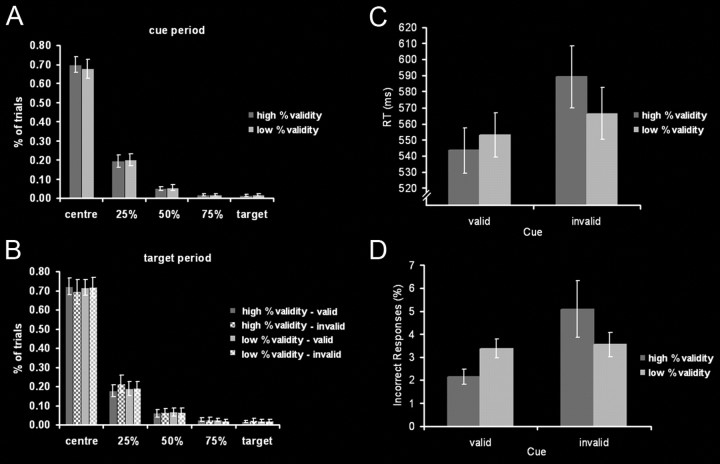
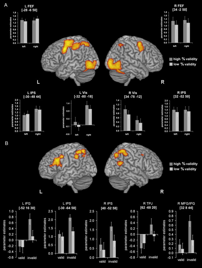
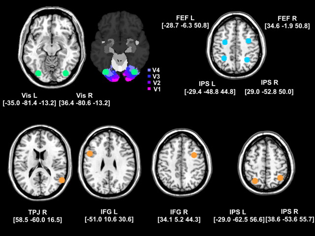
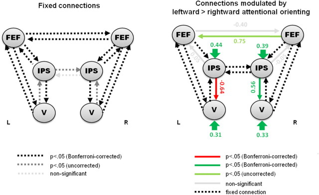
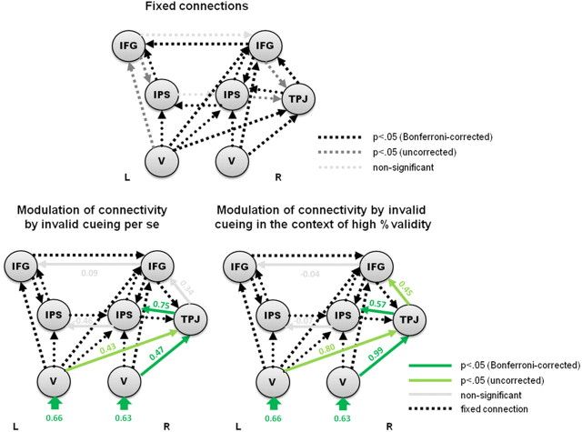
Attentional orientation to a spatial cue and reorientation-after invalid cueing-are mediated by two distinct networks in the human brain. A bilateral dorsal frontoparietal network, comprising the intraparietal sulcus (IPS) and the frontal eye fields (FEF), controls the voluntary deployment of attention and may modulate visual cortex in preparation for upcoming stimulation. In contrast, reorienting attention to invalidly cued targets engages a right-lateralized ventral frontoparietal network comprising the temporoparietal junction (TPJ) and ventral frontal cortex. The present fMRI study investigated the functional architecture of these two attentional systems by characterizing effective connectivity during lateralized orienting and reorienting of attention, respectively. Subjects performed a modified version of Posner's location-cueing paradigm. Dynamic causal modeling (DCM) of regional responses in the dorsal and ventral network, identified in a conventional (SPM) whole-brain analysis, was used to compare different functional architectures. Bayesian model selection showed that top-down connections from left and right IPS to left and right visual cortex, respectively, were modulated by the direction of attention. Moreover, model evidence was highest for a model with directed influences from bilateral IPS to FEF, and reciprocal coupling between right and left FEF. Invalid cueing enhanced forward connections from visual areas to right TPJ, and directed influences from right TPJ to right IPS and IFG (inferior frontal gyrus). These findings shed further light on the functional organization of the dorsal and ventral attentional network and support a context-sensitive lateralization in the top-down (backward) mediation of attentional orienting and the bottom-up (forward) effects of invalid cueing.
Figures
Similar articles
-
What is "odd" in Posner's location-cueing paradigm? Neural responses to unexpected location and feature changes compared.J Cogn Neurosci. 2009 Jan;21(1):30-41. doi: 10.1162/jocn.2009.21003. J Cogn Neurosci. 2009. PMID: 18476756
-
Neural correlates of the spatial and expectancy components of endogenous and stimulus-driven orienting of attention in the Posner task.Cereb Cortex. 2010 Jul;20(7):1574-85. doi: 10.1093/cercor/bhp215. Epub 2009 Oct 21. Cereb Cortex. 2010. PMID: 19846472
-
Effective connectivity during feature-based attentional capture: evidence against the attentional reorienting hypothesis of TPJ.Cereb Cortex. 2014 Dec;24(12):3131-41. doi: 10.1093/cercor/bht172. Epub 2013 Jul 3. Cereb Cortex. 2014. PMID: 23825319
-
The role of the lateral intraparietal area in orienting attention and its implications for visual search.Eur J Neurosci. 2011 Jun;33(11):1982-90. doi: 10.1111/j.1460-9568.2011.07700.x. Eur J Neurosci. 2011. PMID: 21645094 Free PMC article. Review.
-
The neural mechanisms of visual selection: the view from neuropsychology.Ann N Y Acad Sci. 2010 Mar;1191:156-81. doi: 10.1111/j.1749-6632.2010.05448.x. Ann N Y Acad Sci. 2010. PMID: 20392280 Review.
Cited by
-
Rethinking the role of the rTPJ in attention and social cognition in light of the opposing domains hypothesis: findings from an ALE-based meta-analysis and resting-state functional connectivity.Front Hum Neurosci. 2013 Jul 10;7:323. doi: 10.3389/fnhum.2013.00323. eCollection 2013. Front Hum Neurosci. 2013. PMID: 23847497 Free PMC article.
-
Brain networks underlying conscious tactile perception of textures as revealed using the velvet hand illusion.Hum Brain Mapp. 2018 Dec;39(12):4787-4801. doi: 10.1002/hbm.24323. Epub 2018 Aug 10. Hum Brain Mapp. 2018. PMID: 30096223 Free PMC article.
-
Modality-spanning deficits in attention-deficit/hyperactivity disorder in functional networks, gray matter, and white matter.J Neurosci. 2014 Dec 10;34(50):16555-66. doi: 10.1523/JNEUROSCI.3156-14.2014. J Neurosci. 2014. PMID: 25505309 Free PMC article.
-
A distributed effects perspective of dimensionally defined psychiatric disorders: and convergent versus core deficit effects in ADHD.Front Psychiatry. 2014 Jun 5;5:62. doi: 10.3389/fpsyt.2014.00062. eCollection 2014. Front Psychiatry. 2014. PMID: 24926271 Free PMC article. Review.
-
Visuospatial processing: A review from basic to current concepts.Dement Neuropsychol. 2014 Apr-Jun;8(2):175-181. doi: 10.1590/S1980-57642014DN82000014. Dement Neuropsychol. 2014. PMID: 29213900 Free PMC article.
References
-
- Corbetta M, Shulman GL. Control of goal-directed and stimulus-driven attention in the brain. Nat Rev Neurosci. 2002;3:201–215. - PubMed
-
- Corbetta M, Kincade JM, Ollinger JM, McAvoy MP, Shulman GL. Voluntary orienting is dissociated from target detection in human posterior parietal cortex. Nat Neurosci. 2000;3:292–297. - PubMed
Publication types
MeSH terms
Substances
Grants and funding
LinkOut - more resources
Full Text Sources