. 2012 Aug 1;32(31):10662-73.
doi: 10.1523/JNEUROSCI.5785-10.2012.

The stat3/socs3a pathway is a key regulator of hair cell regeneration in zebrafish. [corrected]

Affiliations

The stat3/socs3a pathway is a key regulator of hair cell regeneration in zebrafish. [corrected]

Jin Liang et al. J Neurosci. .

Erratum in

  • J Neurosci. 2012 Oct 3;32(40):14052

Abstract

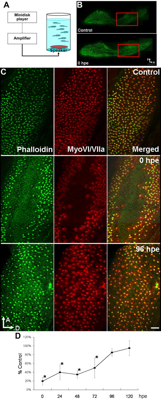
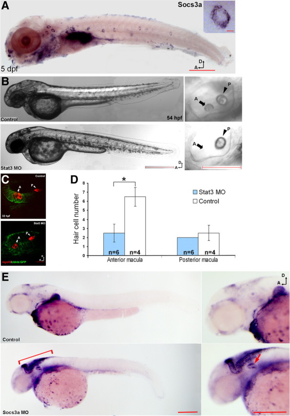
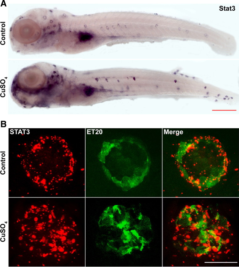
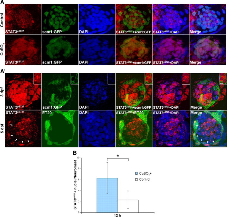
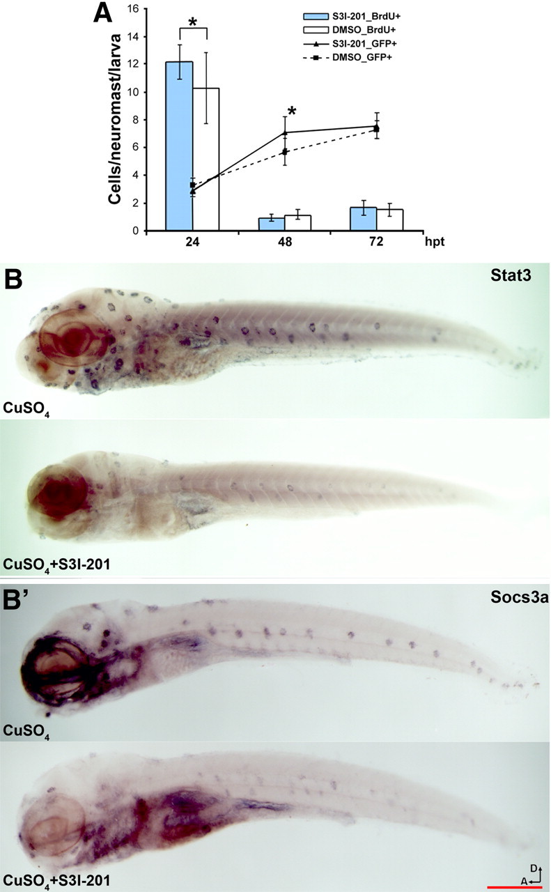
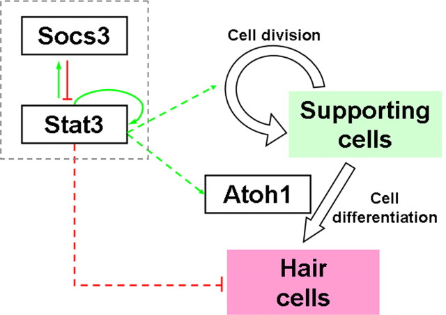
All nonmammalian vertebrates studied can regenerate inner ear mechanosensory receptors (i.e., hair cells) (Corwin and Cotanche, 1988; Lombarte et al., 1993; Baird et al., 1996), but mammals possess only a very limited capacity for regeneration after birth (Roberson and Rubel, 1994). As a result, mammals experience permanent deficiencies in hearing and balance once their inner ear hair cells are lost. The mechanisms of hair cell regeneration are poorly understood. Because the inner ear sensory epithelium is highly conserved in all vertebrates (Fritzsch et al., 2007), we chose to study hair cell regeneration mechanism in adult zebrafish, hoping the results would be transferrable to inducing hair cell regeneration in mammals. We defined the comprehensive network of genes involved in hair cell regeneration in the inner ear of adult zebrafish with the powerful transcriptional profiling technique digital gene expression, which leverages the power of next-generation sequencing ('t Hoen et al., 2008). We also identified a key pathway, stat3/socs3, and demonstrated its role in promoting hair cell regeneration through stem cell activation, cell division, and differentiation. In addition, transient pharmacological inhibition of stat3 signaling accelerated hair cell regeneration without overproducing cells. Taking other published datasets into account (Sano et al., 1999; Schebesta et al., 2006; Dierssen et al., 2008; Riehle et al., 2008; Zhu et al., 2008; Qin et al., 2009), we propose that the stat3/socs3 pathway is a key response in all tissue regeneration and thus an important therapeutic target for a broad application in tissue repair and injury healing.


Figures

Figure 1.
Figure 1.
Saccular hair cells were eliminated by noise exposure and subsequently regenerated. A, Hair cell loss in the saccule was consistently induced by the noise exposure apparatus. B, Phalloidin staining of the saccular hair cells; hair cell loss occurred in the anterior-medial area of the saccular sensory epithelium (red boxes; scale bar, 100 μm). C, Closer examination of the damaged area with phalloidin (green channel) and anti-myosin VI/VIIa (red channel) staining confirmed the complete elimination of hair cell bodies (middle; scale bar, 20 μm) as well as the repopulation of hair cells to control level by 96 hpe in the damaged area (bottom). D, Quantification of hair cell number after sound exposure (one-way ANOVA and post hoc test, n = 3, *p < 0.05; all error bars in this publication demonstrate SD). D, Dorsal; A, anterior.
Figure 2.
Figure 2.
Analyses and confirmation of tag-profiling results from inner ear tissues collected during hair cell regeneration. A, A total number of 303,342 unique-sequence tag sequences from inner ear tissues collected at five time points were mapped against transcriptome and genome databases. Approximately 34% of the tag sequences were mapped to one or more known/predicted transcripts and ∼17% to unique loci in genome (without a known transcript), leaving ∼49% of the sequences without transcriptome or genome mapping. Among those tags mapped to known/predicted transcripts, ∼78% were unambiguously mapped. B, Some of the candidate genes (0 hpe) identified by tag profiling were confirmed by qRT-PCR using GAPDH as a reference gene (n = 3, one-tailed t test, *p = 1.81e-4, 3.78e-4, 9.41e-3, 8.76e-3, 0.0272, 1.578e-4). C, Clustering analysis of the five expression profiles with Genesifter showed the relationship between different profiles (top diagram). The clustering results had the predicted relationship where the 0 hpe expression was the most different from control and then the samples progressively returned to “normal” over time. Pathway analysis showed cell signaling pathways represented by the candidate genes that are critical for specific phases of regeneration [e.g., cell proliferation-related at 0 hpe and cell differentiation-related at later time points (bottom boxes)]. D, Pathway analysis of the candidate genes (identified at 0 hpe) involved in inner ear hair cell regeneration highlighted the interactions between stat3 and socs3 (dashed circles) with other identified candidate genes. The known interactions (red: positive; green: negative; gray: unspecified) between the human orthologs of the candidate genes were extracted in batches to predict the candidate pathways involved in the hair cell regeneration. Different colors of the circles indicate those genes being upregulated (red) or downregulated (blue) during hair cell regeneration.
Figure 3.
Figure 3.
stat3 and socs3a are involved in hair cell production during zebrafish larval development. A, socs3a expression was detected in anterior and posterior lateral line neuromasts in 5 dpf larvae by in situ hybridization; there is a close-up of one neuromast in the top right corner (scale bars: top, 10 μm; bottom, 1 mm). Expression of socs3a is similar to stat3 expression (Oates et al., 1999; Thisse and Thisse, 2004). B, The anterior otolith (arrows labeled with “A”) in stat3 morphants was typically missing, while the posterior otolith (arrowheads labeled with “P”) appeared approximately normal (scale bars: left, 500 μm; right, 100 μm). C, MyosinVI antibody staining (red channel) of the hair cells in the anterior sensory macula (arrows labeled with A) and posterior macula (arrowheads labeled with P) in control larvae and stat3 morphants (scale bar, 20 μm) at 32 hpf. cldnb:GFP (green channel) was used to outline the otic vesicle. D, The stat3 morphants showed a significant reduction in the number of hair cells in the anterior macula, but not in the posterior macula at 32 hpf (n = 4 (control)/6 (MOs), one-tailed t test, *p = 2.79e-4). E, Both stat3 and socs3a morphants possessed fewer posterior lateral line neuromasts (n = 10 in all groups, one-tailed t test, *p = 2.52e-08, 2.41e-04) with a smaller number of hair cells per neuromast (n = 10 in all groups, one-tailed t test, *p = 1.10e-06, 1.56e-10) compared with control larvae at 2.5 dpf. In socs3a morphants (bottom), an expansion of atoh1a expression was detected by in situ hybridization in the brain area (bracket) as well as the otic vesicle (arrow) compared with control (top) (scale bars: left, 1 mm; right, 500 μm). D, Dorsal; A, anterior; MO, morpholino.
Figure 4.
Figure 4.
Hair cell death in the lateral line system resulted in an increase in stat3 expression level in the nonsensory cells in the neuromasts. A, The results of regular whole-mount in situ hybridization targeting stat3 mRNA in control (top) CuSO4-treated (bottom) larvae showed an increase in stat3 expression when hair cell death took place. Scale bar, 1 mm. B, Costaining of neuromasts with fluorescent detection of probes targeting stat3 mRNA (red channel) and FITC-conjugated anti-GFP antibody (green channel) in ET20 transgenic larvae showed an increase in nonsensory cells in the neuromasts after hair cell death. Scale bar, 20 μm.
Figure 5.
Figure 5.
Phosphorylation and nuclear import of stat3 proteins were detected in the regenerating and developing neuromasts. A, Costaining of anti-STAT3pS727 with DAPI in Tg(scm1:GFP) larvae confirmed anti-STAT3pS727 labeling mainly in the nuclei of supporting cells (GFP positive) at 12 hpt with copper compared with untreated controls. Scale bar, 20 μm. A′, In 3 dpf larvae, the majority of the supporting cell nuclei (GFP-positive cells) were labeled with STAT3pS727 antibody (top). Differentiating hair cells showed decreased STAT3pS727 nuclear staining, but an increase of STAT3pS727 labeling in the cytoplasm (top, close-ups; scale bar: close-ups, 5 μm). Nuclear anti-STAT3pS727 labeling decreased dramatically in more mature neuromasts in 5 dpf larvae (bottom), except for a small group of cells labeled with GFP (arrowheads) in ET20 transgenic larvae (scale bars, 20 μm). B, Quantification of activated phospho-STAT3. The increase in nuclear localized phospho-stat3 was statistically significant (n = 10/9, one-tailed t test, p = 9.83e-4). S727 in human STAT3 corresponds to S751 in zebrafish STAT3 by sequence alignment.
Figure 6.
Figure 6.
S3I-201 promoted lateral line hair cell regeneration by downregulating stat3/socs3 signaling. A, Quantification of hair cells after lateral line hair cell loss induced by CuSO4 treatment; larvae incubated with S3I-201 had more hair cells [GFP-positive cells in Tg(pou4f3:GFP)] per neuromast at 48 hpt with CuSO4 compared with the control larvae incubated with DMSO (n = 12/9, one-tailed t test, *p = 5.34e-3), but not at 24 or 72 hpt (line graph). BrdU incorporation assays showed a significantly higher number of BrdU-positive cells per neuromast in larvae incubated with S3I-201 at 24 hpt (n = 16/10; one-tailed t test, *p = 9.24e-3), but not at later time points (bar graph). B, B′, 5 dpf larvae fish were treated with CuSO4 with or without the presence of S3I-201 for 1 h and examined with in situ hybridization. stat3 (B) and socs3a (B′) expression levels were both reduced in larvae treated with both CuSO4 and S3I-201 (B and B′, top) compared with those treated with only CuSO4 (B and B′, bottom). Scale bar, 1 mm. D, Dorsal; A, anterior.
Figure 7.
Figure 7.
The working model of the stat3/socs3 pathway and the interaction of stat3/socs3 with atoh1 during hair cell regeneration in zebrafish. stat3 positively regulates the function of atoh1, a key player in promoting hair cell fate commitment, in a direct or indirect manner. In addition, the balancing of the stat3/socs3 pathway is crucial to hair cell differentiation: either hyperactivation or hypoactivation of stat3 inhibits hair cell differentiation. The self-restrictive loop helps to balance the expression of stat3 spatially and temporally to ensure the appropriate replenishment of the lost hair cells.

Similar articles

Cited by

References

    1. Amsterdam A, Burgess S, Golling G, Chen W, Sun Z, Townsend K, Farrington S, Haldi M, Hopkins N. A large-scale insertional mutagenesis screen in zebrafish. Gene Dev. 1999;13:2713–2724. - PMC - PubMed
    1. Baird RA, Steyger PS, Schuff NR. Mitotic and nonmitotic hair cell regeneration in the bullfrog vestibular otolith organs. Ann NY Acad Sci. 1996;781:59–70. - PubMed
    1. Balak KJ, Corwin JT, Jones JE. Regenerated hair cells can originate from supporting cell progeny: evidence from phototoxicity and laser ablation experiments in the lateral line system. J Neurosci. 1990;10:2502–2512. - PMC - PubMed
    1. Bang PI, Sewell WF, Malicki JJ. Morphology and cell type heterogeneities of the inner ear epithelia in adult and juvenile zebrafish (Danio rerio) J Comp Neurol. 2001;438:173–190. - PubMed
    1. Behra M, Bradsher J, Sougrat R, Gallardo V, Allende ML, Burgess SM. Phoenix is required for mechanosensory hair cell regeneration in the zebrafish lateral line. PLoS Genet. 2009;5:e1000455. - PMC - PubMed

Publication types

MeSH terms