Review
. 2012 Aug 2;14(1):54.
doi: 10.1186/1532-429X-14-54.

Diagnostic and prognostic value of cardiovascular magnetic resonance in non-ischaemic cardiomyopathies

Affiliations
Review

Diagnostic and prognostic value of cardiovascular magnetic resonance in non-ischaemic cardiomyopathies

Chirine Parsai et al. J Cardiovasc Magn Reson. .

Abstract

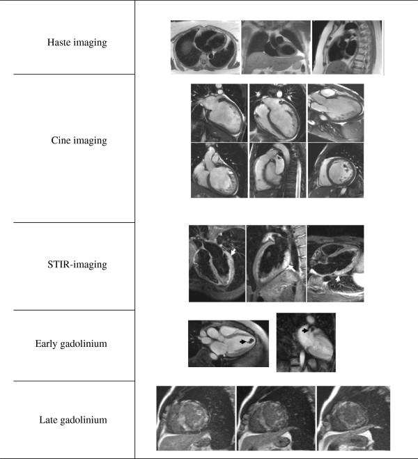
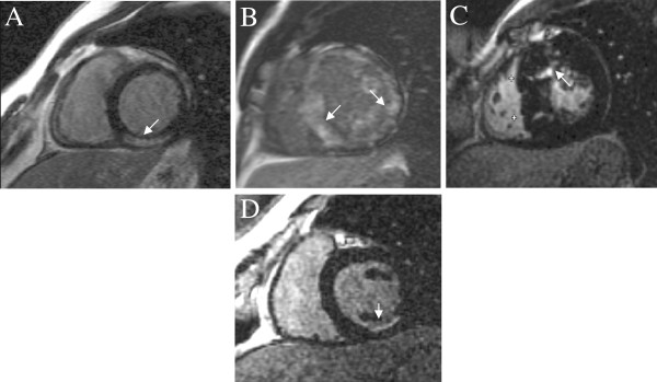
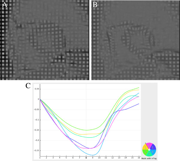
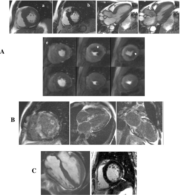
Cardiovascular Magnetic Resonance (CMR) is recognised as a valuable clinical tool which in a single scan setting can assess ventricular volumes and function, myocardial fibrosis, iron loading, flow quantification, tissue characterisation and myocardial perfusion imaging. The advent of CMR using extrinsic and intrinsic contrast-enhanced protocols for tissue characterisation have dramatically changed the non-invasive work-up of patients with suspected or known cardiomyopathy. Although the technique initially focused on the in vivo identification of myocardial necrosis through the late gadolinium enhancement (LGE) technique, recent work highlighted the ability of CMR to provide more detailed in vivo tissue characterisation to help establish a differential diagnosis of the underlying aetiology, to exclude an ischaemic substrate and to provide important prognostic markers. The potential application of CMR in the clinical approach of a patient with suspected non-ischaemic cardiomyopathy is discussed in this review.


Figures

Figure 1
Figure 1
Standard cardiomyopathy protocol. Standard cardiomyopathy protocol including HASTE, cine imaging, T2-weighted-imaging (STIR) searching for inflammation/edema (white arrows), early gadolinium imaging to detect thrombus (black arrow) and LGE to identify fibrosis or infiltration (diffuse LGE typical of amyloidosis).
Figure 2
Figure 2
Diagnostic patterns of LGE. Distribution pattern and location of LGE contributes to the differential diagnosis between a NICMP (A : sub-epicardial fibrosis following myocarditis, B : circumferential diffuse enhancement in amyloidosis, C : patchy fibrosis in affected hypertrophied segments in HCM) and a typical ischaemic sub-endocardial enhancement (D).
Figure 3
Figure 3
Example of myocardial tagging using CSPAMM in an HCM patient (A: end diastole, B. end systole). Deformation curves are extracted using a dedicated software (C). Courtesy of Dr Tevfik Ismael. Images were analysed with inTag (software supplied as part of a collaboration agreement with University of Lyon, Pierre Croisille and Patrick Clarysse, Creatis).
Figure 4
Figure 4
Thickened myocardium. (A) HCM with asymmetrical septal hypertrophy (a, arrow) and a confluent LGE pattern matching the hypertrophy (b, arrow). Dynamic LVOT obstruction with SAM (c: diastole, d: systole with a SAM) and circumferential sub-endocardial ischemia in hypertrophied areas as a consequence of microvascular dysfunction are among classical features (e top: stress-induced perfusion defects, e bottom: normalization at rest). (B) Amyloidosis with a typical circumferential diffuse enhancement (‘zebra’ pattern). (C) Anderson-Fabry disease with concentric LV hypertrophy (a) and patchy LGE affecting the inferolateral wall (b, arrow).
Figure 5
Figure 5
Pattern of fibrosis in DCM. Typical mid-wall LGE seen in DCM (A, arrows) differing from the sub-endocardial ischaemic pattern (B).
Figure 6
Figure 6
ARVC. Findings suggestive of ARVC include localized aneurysms in the RV outflow tract (arrows, A: diastole, B: systole), LGE of the free wall (C, arrow) and in the left-sided form areas of mid-wall LGE at the RV-LV insertion point (D, arrow).
Figure 7
Figure 7
Acute chest pain syndromes (A) Acute viral myocarditis displaying edema on STIR images (a-b, arrows) and typical sub-epicardial LGE (c-d, arrows). (B) Tako-tsubo with acute apical ballooning of the LV (a: diastole, b: systole displaying apical akinesia, arrows) without LGE (c) followed by complete LV functional recovery at follow-up (d: diastole, e: systole).
Figure 8
Figure 8
Sarcoidosis. Hilar lymphadenopathy on HASTE (A, arrows), localized edema in the lateral wall on STIR images (B, arrow) and mid-wall LGE (C, arrow) are suggestive of sarcoidosis.
Figure 9
Figure 9
Left ventricular noncompaction LVNC with marked LV apical and lateral wall trabeculations, arrows (left panel: diastole, right panel: systole, A: mid-segment, B: apical segment, C: 2-chamber view).
Figure 10
Figure 10
Myocardial iron overload. A mid-level gradient echo short-axis slice is imaged at different echo times (A: 2 ms to D: 14 ms) and T2*value is estimated from the signal intensity decay curve (E). Typical sub-epicardial iron deposition can be seen (arrow).

References

    1. Maron BJ, Towbin JA, Thiene G, Antzelevitch C, Corrado D, Arnett D, Moss AJ, Seidman CE, Young JB. Contemporary definitions and classification of the cardiomyopathies: an American Heart Association Scientific Statement from the Council on Clinical Cardiology, Heart Failure and Transplantation Committee; Quality of Care and Outcomes Research and Functional Genomics and Translational Biology Interdisciplinary Working Groups; and Council on Epidemiology and Prevention. Circulation. 2006;113:1807–1816. doi: 10.1161/CIRCULATIONAHA.106.174287. - DOI - PubMed
    1. Aletras AH, Kellman P, Derbyshire JA, Arai AE. ACUT2E TSE-SSFP: a hybrid method for T2-weighted imaging of edema in the heart. Magn Reson Med. 2008;59:229–235. doi: 10.1002/mrm.21490. - DOI - PubMed
    1. Elliott P, Andersson B, Arbustini E, Bilinska Z, Cecchi F, Charron P, Dubourg O, Kuhl U, Maisch B, McKenna WJ. et al.Classification of the cardiomyopathies: a position statement from the European Society Of Cardiology Working Group on Myocardial and Pericardial Diseases. Eur Heart J. 2008;29:270–276. - PubMed
    1. Assomull RG, Shakespeare C, Kalra PR, Lloyd G, Gulati A, Strange J, Bradlow WM, Lyne J, Keegan J, Poole-Wilson P. et al.Role of Cardiovascular Magnetic Resonance as a Gatekeeper to Invasive Coronary Angiography in Patients Presenting With Heart Failure of Unknown Etiology. Circulation. 2011;124:1351–1360. doi: 10.1161/CIRCULATIONAHA.110.011346. - DOI - PubMed
    1. Kramer CM, Barkhausen J, Flamm SD, Kim RJ, Nagel E. Standardized cardiovascular magnetic resonance imaging (CMR) protocols, society for cardiovascular magnetic resonance: board of trustees task force on standardized protocols. Journal of Cardiovascular Magnetic Resonance. 2008;10:35. doi: 10.1186/1532-429X-10-35. - DOI - PMC - PubMed

Publication types