. 2012 Sep 14;47(5):707-21.
doi: 10.1016/j.molcel.2012.06.025. Epub 2012 Aug 1.

Molecular basis for specific regulation of neuronal kinesin-3 motors by doublecortin family proteins

Affiliations

Molecular basis for specific regulation of neuronal kinesin-3 motors by doublecortin family proteins

Judy S Liu et al. Mol Cell. .

Abstract

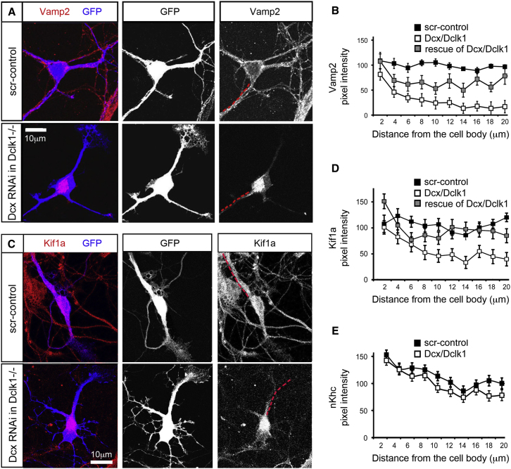
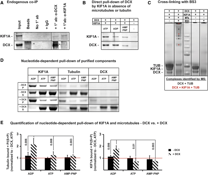
Doublecortin (Dcx) defines a growing family of microtubule (MT)-associated proteins (MAPs) involved in neuronal migration and process outgrowth. We show that Dcx is essential for the function of Kif1a, a kinesin-3 motor protein that traffics synaptic vesicles. Neurons lacking Dcx and/or its structurally conserved paralogue, doublecortin-like kinase 1 (Dclk1), show impaired Kif1a-mediated transport of Vamp2, a cargo of Kif1a, with decreased run length. Human disease-associated mutations in Dcx's linker sequence (e.g., W146C, K174E) alter Kif1a/Vamp2 transport by disrupting Dcx/Kif1a interactions without affecting Dcx MT binding. Dcx specifically enhances binding of the ADP-bound Kif1a motor domain to MTs. Cryo-electron microscopy and subnanometer-resolution image reconstruction reveal the kinesin-dependent conformational variability of MT-bound Dcx and suggest a model for MAP-motor crosstalk on MTs. Alteration of kinesin run length by MAPs represents a previously undiscovered mode of control of kinesin transport and provides a mechanism for regulation of MT-based transport by local signals.


Figures

Figure 1
Figure 1
Kif1a Is Mislocalized in Dcx/Dclk1-Deficient Neurons (A) Dissociated WT and Dclk1−/− hippocampal neurons are transfected with either a scrambled control shRNAi or a Dcx shRNAi plasmid with a GFP reporter and immunostained for Vamp2 after 4 DIV. (B) Quantification of Vamp2 intensity along the trajectories of neural processes starting from the soma and extending out 20 μm (shown as a broken red line adjacent to the neurite in A) demonstrates statistically significantly (p < 0.05) lower levels of Vamp2 starting at 4 μm from the cell-body Dcx/Dclk- deficient neurons (n = 32) as compared with control (n = 29) in one representative experiment out of four. The Vamp2 level in neurites is partially restored by expression of the shRNAi-resistant HA-Dcx (p < 0.05, n = 23). (C) WT or Dclk1−/− neurons are transfected with a plasmid for GFP expression to mark neuronal morphology and shRNAi specific for Dcx where indicated. (D) The pixel intensity of Kif1a versus distance from the cell body of the neuron is shown for WT and Dcx RNAi-treated Dclk1−/− neurons, demonstrating significantly less Kif1a neurites of Dcx/Dclk1-deficient neurons after 4 μm from the cell body, and is partially restored by rescue by overexpression of Dcx (n = 37, 31, and 25, respectively). (E) The pixel intensity of neuronal kinesin heavy chain (nKhc) versus distance from the cell body of the neuron is shown for WT and Dcx shRNAi-treated Dclk1−/− neurons demonstrating no change in Dcx/Dclk1-deficient neurons (n = 30 and 32, respectively). Error bars represent the standard error of the mean (SEM). Scale bars in all panels represent 10 μm.
Figure 2
Figure 2
Vamp2 Transport from the Cell Body into Neurites Is Dependent on Kif1a (A)–(C) show DIV4 neurons with high power views (middle/bottom panels) of neurites with both Vamp2 immunostaining (red) and GFP (green), a marker of successful transfection with the shRNAi construct. (A) WT neurons are transfected with a scrambled control showing Vamp2 in neurite of the green cell (white arrows). (B) Knockdown of Kif1a by RNAi results in a majority of neurons with Vamp2 only in the cell body. High-power views (middle/bottom panels) show lack of Vamp2 in the neurites (white arrows). (C) Kif1a knockdown neurons where Vamp2 is clumped in the neurites (white arrows). (D) Quantification of Vamp2-GFP vesicles that moved more than three microns over 120 s are shown for control (56%), knockdown neurons (4%), and rescue (37%). Error bars represent SEM. (E–G) Top panels are the first frame of a 120 s time-lapse video of Vamp2-GFP in control, Kif1a knockdown neurons, and rescue neurons. Bottom panels are the tracking of the Vamp2-GFP. Each color represents the track of a single Vamp2 vesicle over the full 120 s. (H–M) (H) Colocalization of Vamp2-RFP and Kif1a-mCitrine is shown in a DIV5 WT neuron. (I) depicts the Vamp2-RFP channel and (J) the Kif1a-mCitrine channel. (K) Minimal colocalization of Mito-RFP and Kif1a-mCitrine is shown in a DIV5 WT neuron with (L) depicting Mito-RFP and (M) Kif1a-mCitrine. Scale bars, 10 μm.
Figure 3
Figure 3
Efflux of Vamp2-GFP into Neurites Is Impaired in Dcx/Dclk1-Deficient Neurons (A–D) Shown is live-cell imaging of control, Dcx-deficient, Dcx/Dclk1-deficient, and HA-Dcx rescue of Dcx/Dclk1 deficient neurons. In (A)–(D), the top left panel is the first frame of the imaging study. A red, broken line 10 μm in length shows the region of the neurite used for generating the kymograph in the bottom left panel. The kymograph is created using the pixels selected by the tracing of the neurite from point A to point B. A red line on the left marks the 28 s time interval depicted by frames in the panel on the right. The right panels show frames at 7 s intervals of the 10 μm region of interest. White arrows mark the position of Vamp2-GFP transport packets. Scale bars, 10 μm in all panels. (E) Quantification of efflux (vesicle exit of the cell body into the neurite) is shown for control, Dcx RNAi, Dcx RNAi in Dclk1−/− neurons and Dcx RNAi in Dclk1−/− with rescue by expression of HA-Dcx. (F) A determination of the number of Vamp2-GFP transport packets seen in (A)–(D) shows a significant decrease in the number of Vamp2-GFP vesicles in the Dcx/Dclk1 double deficient neurons, which can be rescued by expression of HA-Dcx. Error bars in (E) and (F) respresent SEM.
Figure 4
Figure 4
The Run Length of Vamp2-GFP in Dcx/Dclk1-Deficient Neurons Is Decreased (A–C) WT neurons treated with a scrambled control (A) and Dcx shRNAi (B) are then transfected with a plasmid for expression of Vamp2-GFP for live imaging. The top panel shows the first frame, and the bottom panel shows the tracks of Vamp2-GFP transport packets within the neurites. Each color represents the track of a single Vamp2 vesicle over the full 120 s. (C) Vamp2-GFP vesicles were analyzed for number of mobile vesicles in Dcx RNAi-treated neurons, Dcx/Dclk1 double deficient neurons, and rescue conditions. (D) Average run lengths are shown for each condition. This analysis excluded Vamp2-GFP vesicles that moved less than 3 μm in 120 s, as these may reflect vesicles in which the necessary components (e.g., MT, motor, cargo) are not properly complexed. (E) Velocity is shown in Dcx/ Dclk1-deficient, rescue, or overexpression conditions. (F–J) Mitochondrial transport is imaged in control neurons (F) and Dcx shRNAi neurons (G) using transfection with Mito-DsRed. The top panel shows the first frame, and the bottom panel shows the tracks of Mito-DsRed within the neurites over 120 s. Mitochondrial transport in neurites does not change significantly in terms of percent mobile organelles (H), run length (I), and velocity (J). Error bars in all panels represent the SEM. Scale bar, 5 μm in all panels.
Figure 5
Figure 5
Causative Mutations for Lissencephaly Alter Kif1a/Vamp2 Transport (A) Dcx binding to MTs in normal neurons is shown. (B and C) Neurons are transfected with Dcx shRNAi and rescued with HA-tagged WT or mutant Dcx constructs resistant to the shRNAi. (B) depicts the distribution of WT HA-Dcx, which is similar to that of endogenous Dcx with more Dcx in the distal neurites, albeit higher levels of Dcx overall. (C) Mutant Dcx S47R binds only in the cell body. (D–G) Vamp2-GFP transport out of the cell body into neurites is shown in Dcx shRNAi neurons rescued by either WT HA-Dcx or HA-Dcx S47R. (D and E) Both efflux of Vamp2-GFP and number of Vamp2-GFP vesicles in neurites are shown for rescue with either WT HA-DcxS47R. (F and G) The top left panel is the first frame of the imaging study. A red, broken line 10 μm in length shows the region of the neurite used for generating the kymograph in the bottom left panel. The kymograph is created using the pixels selected by the tracing of the neurite from point A to point B. These pixels are aligned sequentially from the first frame to the last frame so that vesicle movement in the region of interest is shown throughout the imaging study. A red line on the left marks the 28 s time interval depicted by frames in the panel on the right. The right panels of (F) and (G) show frames of the neurite used to generate the kymograph at 7 s intervals. (H) The Dcx mutation W146C does not affect the MT binding of Dcx. (I) Top panels show the first frame of the time-lapse sequences used to generate the Vamp2-GFP tracks shown in the bottom panel for rescue with either WT HA-Dcx or HA-Dcx W146C. (J) Numbers of mobile vesicles, run length, and velocity are quantified in the WT HA-Dcx and HA-Dcx W146C rescue conditions. Error bars in all panels represent the SEM.
Figure 6
Figure 6
Dcx Interacts with Kif1a and Facilitates Binding of the Low-Affinity, ADP-Bound Kif1a Motor to MTs (A) Coimmunoprecipitation of endogenous Dcx and Kif1a from human fetal cortex (23 weeks) was performed with antisera to Dcx and Kif1a, respectively, in 2 mM AMP-PNP using BSA-blocked protein G beads. Protein complexes were analyzed by western blot. Lane 1 shows the original protein lysate at a 1:20 dilution. Lanes 2–4 are negative controls: (2) blocked protein G beads without lysate, (3) beads incubated with lysate but without antibody, (4) beads incubated with lysate and a nonspecific IgG antibody. Lane 5 shows pull-down of Kif1a with the primary polyclonal Dcx antibody; the Kif1a band is clearly visible. Lane 6 shows pull-down of Kif1a with the primary Kif1a antibody, but very little Dcx coimmunoprecipitates (faint band marked by asterisk). (B) Direct pull-down of overexpressed and purified full-length human Dcx by an N-terminal HaloTag human Kif1a (amino acids 1–361) fusion protein was performed in the presence of 4 mM nucleotides and 5 μM of each protein using HaloLink magnetic beads. Lanes 1–3 show that Dcx and the motor domain of Kif1a interact independently in the absence of MTs and the presence of either ATP, ADP, or AMP-PNP; the presence of excess Kif1a in the pull-down further suggests the existence of more than one binding site of the kinesin-3 motor domain on Dcx-decorated MTs. Lane 4 is a negative control. (C) Dcx and Kif1a form a ternary complex on the MT. Crosslinking was performed using BS3-d0 with purified human Dcx, Kif1a, and porcine MTs as shown. A range of crosslinked Dcx:MT:Kif1a complexes was identified as indicated by the red asterisks in lane 2. Crosslinking in the absence of MTs (lane 4) did not yield any visible bands. (D) Nucleotide-dependent pull-down of MTs in the presence and absence of full-length human Dcx by an N-terminal HaloTag human Kif1a (amino acids 1–361) fusion protein was performed in presence of 4 mM nucleotides and 5 μM of each protein component using HaloLink magnetic beads. Supernatant and pellet fractions are shown to indicate equal total protein loading for each nucleotide condition, and both fractions were used to quantify band intensities by densitometry after silver staining. (E) Quantification of (D) shows that Kif1a binding to MTs in the presence of Dcx and 4 μM nucleotide is significantly enhanced by addition of ADP, but not ATP or AMP-PNP (left panel) when compared to binding in absence of Dcx. Similarly, Dcx enhances pull-down of excess Kif1a motor domain in the ADP binding state, but not in the ATP or AMP-PNP binding state (right panel). Bound fractions were calculated as P/(S+P), and all quantifications are normalized to ATP in absence of DCX as indicated by the red line across all graphs. Error bars represent standard deviation, and significant p values are shown (two-tailed t test, n ≥ 3).
Figure 7
Figure 7
Model of Dcx MT Binding in the Presence and Absence of Kif1a (A) Cryo-EM structures of Dcx-MTs alone (top panel, 8.3 Å resolution, see Figure S5A) and in the presence of the kinesin motor domain (bottom panel; Fourniol et al., 2010); transparent surface, tubulin colored in gray, kinesin in faded pink, Dcx in yellow, docked with atomic coordinates (ribbons) of tubulin (2XRP.PDB, alpha in blue, beta in cyan, crosscorrelation of the fit; top panel, 0.720; bottom panel, 0.744). Kinesin binding affects the structure/flexibility of the linker regions N and C terminal of N-DC (residues at boundaries are numbered). Kif1a loops L2 and L8 (bright pink) are likely in direct contact with linker regions. In the absence of kinesin (N-DC colored yellow), extra density in the reconstruction enabled modeling of the C-terminal linker docked against N-DC through W146 and in good agreement with NMR studies (crosscorrelation 0.684 before and 0.739 after modeling; Kim et al., 2003; also see Figure S5B). The plus end of the MT is oriented upward. (B) Resolution 8.2 Å cryo-electron microscopy reconstruction of Dcx-MTs codecorated with conventional kinesin motor domain (Fourniol et al., 2010; also see Figure S5C), Dcx R1 (1MJD.PDB, model 11, amino acids 46–139, orange, crosscorrelation 0.722) and KIF1A (1I5S.PDB, dark pink, crosscorrelation 0.711). The bound DC domain is surrounded by four motor domains (labeled I–IV), and is <5 Å from motor domains II and III: residues in Kif1A and N-DC separated by less than 5 Å are listed in the table. (C) The superimposition of pseudoatomic models of MT-bound Dcx in the presence (orange ribbon) and absence (yellow ribbon) of Kif1a motor domain (dark pink) shows a clash between the C-terminal linker of N-DC (arrow, thicker yellow ribbon) and Kif1a, explaining why that linker is induced to undock upon motor binding (also see Movie S6).

References

    1. Bai J., Ramos R.L., Ackman J.B., Thomas A.M., Lee R.V., LoTurco J.J. RNAi reveals doublecortin is required for radial migration in rat neocortex. Nat. Neurosci. 2003;6:1277–1283. - PubMed
    1. Bielas S.L., Serneo F.F., Chechlacz M., Deerinck T.J., Perkins G.A., Allen P.B., Ellisman M.H., Gleeson J.G. Spinophilin facilitates dephosphorylation of doublecortin by PP1 to mediate microtubule bundling at the axonal wrist. Cell. 2007;129:579–591. - PMC - PubMed
    1. Bilimoria P.M., de la Torre-Ubieta L., Ikeuchi Y., Becker E.B., Reiner O., Bonni A. A JIP3-regulated GSK3beta/DCX signaling pathway restricts axon branching. J. Neurosci. 2010;30:16766–16776. - PMC - PubMed
    1. Binder L.I., Frankfurter A., Rebhun L.I. Differential localization of MAP-2 and tau in mammalian neurons in situ. Ann. N Y Acad. Sci. 1986;466:145–166. - PubMed
    1. Caviston J.P., Holzbaur E.L. Microtubule motors at the intersection of trafficking and transport. Trends Cell Biol. 2006;16:530–537. - PubMed

Publication types

LinkOut - more resources