Review
. 2012 Oct;181(4):1126-41.
doi: 10.1016/j.ajpath.2012.06.030. Epub 2012 Aug 2.

Mechanisms of glioma-associated neovascularization

Affiliations
Review

Mechanisms of glioma-associated neovascularization

Matthew E Hardee et al. Am J Pathol. 2012 Oct.

Abstract

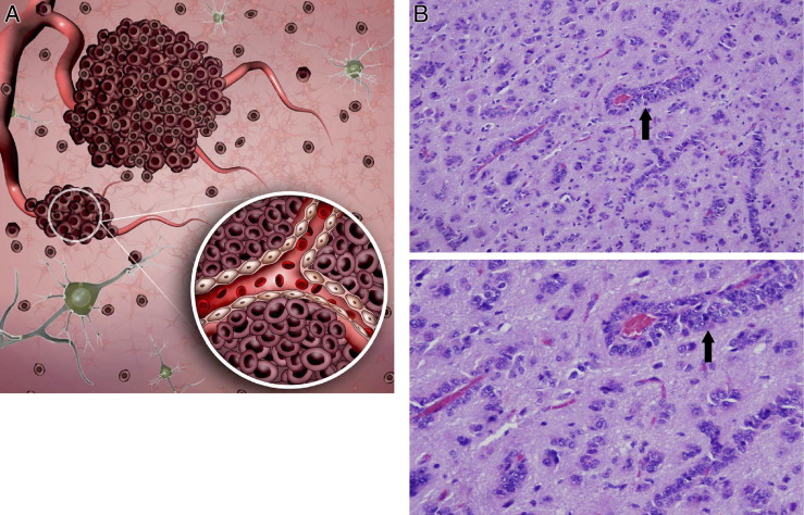
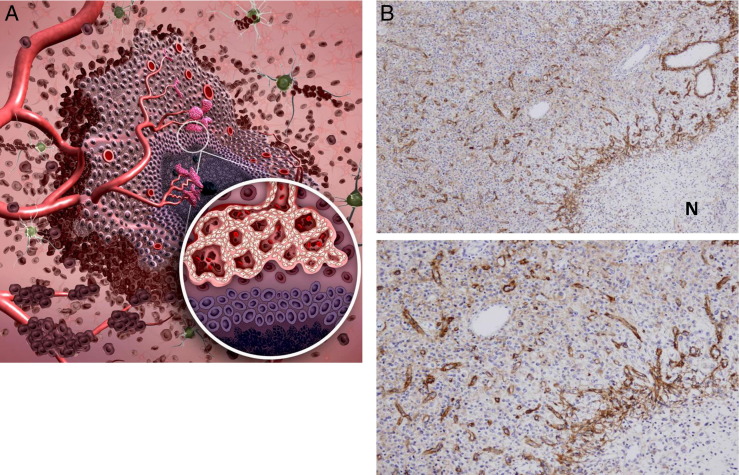
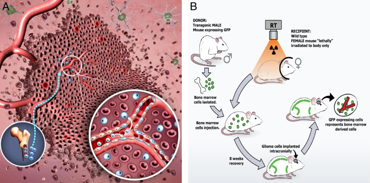
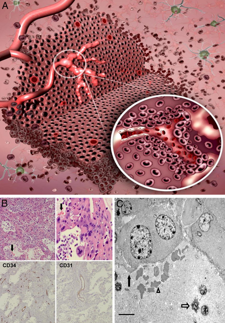
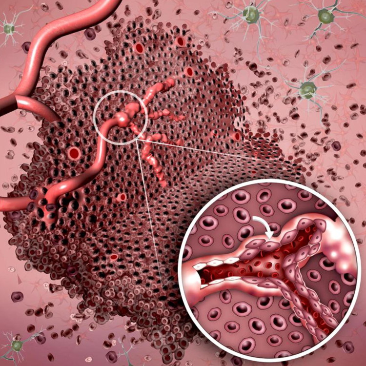
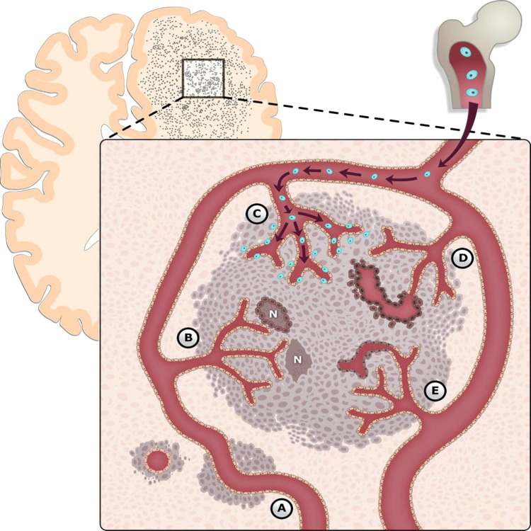
Glioblastomas (GBMs), the most common primary brain tumor in adults, are characterized by resistance to chemotherapy and radiotherapy. One of the defining characteristics of GBM is an abundant and aberrant vasculature. The processes of vascular co-option, angiogenesis, and vasculogenesis in gliomas have been extensively described. Recently, however, it has become clear that these three processes are not the only mechanisms by which neovascularization occurs in gliomas. Furthermore, it seems that these processes interact extensively, with potential overlap among them. At least five mechanisms by which gliomas achieve neovascularization have been described: vascular co-option, angiogenesis, vasculogenesis, vascular mimicry, and (the most recently described) glioblastoma-endothelial cell transdifferentiation. We review these mechanisms in glioma neovascularization, with a particular emphasis on the roles of hypoxia and glioma stem cells in each process. Although some of these processes are well established, others have been identified only recently and will need to be further investigated for complete validation. We also review strategies to target glioma neovascularization and the development of resistance to these therapeutic strategies. Finally, we describe how these complex processes interlink and overlap. A thorough understanding of the contributing molecular processes that control the five modalities reviewed here should help resolve the treatment resistance that characterizes GBMs.


Figures

Figure 1
Figure 1
Vascular co-option. A: Temporally, vascular co-option is the first process by which gliomas attain a vascular supply. The process involves organization of tumor cells into cuffs around normal microvessels (inset). Vascular co-option has been shown to precede angiogenesis in tumor models by up to 4 weeks. B: Photomicrographs of tumor cells in a sectioned human GBM specimen stained with H&E show vascular co-option as classic perivascular cuffs. Original magnification: ×50 (top); ×200 (bottom).
Figure 2
Figure 2
Angiogenesis. A: Angiogenesis follows vascular co-option during tumor vasculature development and is defined as the development of new vessels from pre-existing ones. Hypoxic pseudopalisading glioma cells around necrosis (inset) release proangiogenic factors. This results in the shift of the angiogenic balance toward a proangiogenic phenotype, inducing sprouting from pre-existing vessels. Hypoxia-independent mechanisms driving angiogenesis have also been described. B: Photomicrographs of a sectioned human GBM specimen stained for tenascin-C shows sprouting angiogenesis. Note the gradient from relatively poorly vascularized regions with little angiogenesis (upper left in each panel) to highly vascularized regions with hyperplastic vessels around necrosis (N) (lower right in each panel). Original magnification: ×20 (top); ×100 (bottom).
Figure 3
Figure 3
Vasculogenesis. A: Vasculogenesis involves the mobilization, differentiation, and recruitment of BMDCs (blue). Similar to the angiogenic switch, vasculogenesis is induced largely by hypoxia-mediated release of various factors, the most studied of which are involved in the SDF-1α/CXCR4 pathway. Circulating precursor cells are not only incorporated into the neovasculature, but also can enter the tumor and become tumor-associated macrophages (inset). B: A transgenic murine model is used in our laboratory to study vasculogenesis in GL261 gliomas. When bone marrow cells from a donor male transgenic mouse are transplanted into a recipient female wild-type mouse, marrow-derived cells will express the Y chromosome and marrow-derived EPCs will express GFP detectable by fluorescence microscopy, immunohistochemistry, and flow cytometry. These marrow transplant mice can be implanted intracranially 8 weeks later with syngeneic GL261 glioma cells. Any contribution of GFP+ transplanted BMDCs from the male donor to the endothelium and/or intratumoral cellular milieu is easily detectable.
Figure 4
Figure 4
Vascular mimicry. A: Vascular mimicry is the ability of tumor cells to form functional, perfused, vessel-like networks. These tumor cells lining the vascular channels maintain glioma morphological characteristics and typical glioma markers (inset). B: Photomicrographs demonstrating vascular mimicry. Top row: Low- and high-power views of H&E-stained sections of a human GBM demonstrate perfused vascular networks containing red blood cells (arrows). Bottom row: Immunohistochemical staining of this tumor with CD34 (left, immunoperoxidase) and CD31 (right, immunoperoxidase) reveals immunoreactive vascular channels different from those lined by tumor cells and confirms that these channels are not lined by endothelial cells. C: Electron micrograph shows tumor cells (black arrow) lining a vascular channel containing red blood cells (outlined arrowhead) and polymorphonuclear leukocytes (outlined arrow) in a sectioned human GBM. Scale bar = 10 μm. Original magnification: ×50 (B, top left, bottom left, and bottom right); ×200 (B, top right).
Figure 5
Figure 5
Glioblastoma-endothelial cell transdifferentiation. The mechanism of glioma neovascularization involves transdifferentiation of glioma cells into endothelial cells lining vascular channels (inset), including both a phenotypic transformation and the expression of typical endothelial-specific markers.
Figure 6
Figure 6
Mechanisms of glioma-associated neovascularization. Glioma neovascularization is a complex and highly regulated process, dependent on the balance of at least five separate pathways: A, vascular co-option; B, angiogenesis; C, vasculogenesis; D, vascular mimicry; and E, glioblastoma-endothelial cell transdifferentiation. These mechanisms are likely intimately connected, with perturbations in one pathway altering the vascular phenotype. Although there does appear to be a temporal framework by which gliomas attain vasculature, it is likely that several of these processes can and do occur simultaneously in different microdomains of the tumor. BMDCs are shown in blue. N, necrosis.

References

    1. Brem S., Cotran R., Folkman J. Tumor angiogenesis: a quantitative method for histologic grading. J Natl Cancer Inst. 1972;48:347–356. - PubMed
    1. Heddleston J.M., Li Z., Lathia J.D., Bao S., Hjelmeland A.B., Rich J.N. Hypoxia inducible factors in cancer stem cells. Br J Cancer. 2010;102:789–795. - PMC - PubMed
    1. Semenza G.L. Defining the role of hypoxia-inducible factor 1 in cancer biology and therapeutics. Oncogene. 2010;29:625–634. - PMC - PubMed
    1. Heddleston J.M., Li Z., McLendon R.E., Hjelmeland A.B., Rich J.N. The hypoxic microenvironment maintains glioblastoma stem cells and promotes reprogramming towards a cancer stem cell phenotype. Cell Cycle. 2009;8:3274–3284. - PMC - PubMed
    1. Holash J., Maisonpierre P.C., Compton D., Boland P., Alexander C.R., Zagzag D., Yancopoulos G.D., Wiegand S.J. Vessel cooption, regression, and growth in tumors mediated by angiopoietins and VEGF. Science. 1999;284:1994–1998. - PubMed

Publication types

MeSH terms

Substances