

Review
doi: 10.1038/cdd.2012.96.
Epub 2012 Aug 3.
MicroRNAs tune cerebral cortical neurogenesis
Affiliations
- PMID: 22858543
- PMCID: PMC3438503
- DOI: 10.1038/cdd.2012.96
Item in Clipboard
Review
MicroRNAs tune cerebral cortical neurogenesis
Cell Death Differ.
2012 Oct.
Abstract
MicroRNAs (miRNAs) are non-coding RNAs that promote post-transcriptional silencing of genes involved in a wide range of developmental and pathological processes. It is estimated that most protein-coding genes harbor miRNA recognition sequences in their 3' untranslated region and are thus putative targets. While functions of miRNAs have been extensively characterized in various tissues, their multiple contributions to cerebral cortical development are just beginning to be unveiled. This review aims to outline the evidence collected to date demonstrating a role for miRNAs in cerebral corticogenesis with a particular emphasis on pathways that control the birth and maturation of functional excitatory projection neurons.
Figures
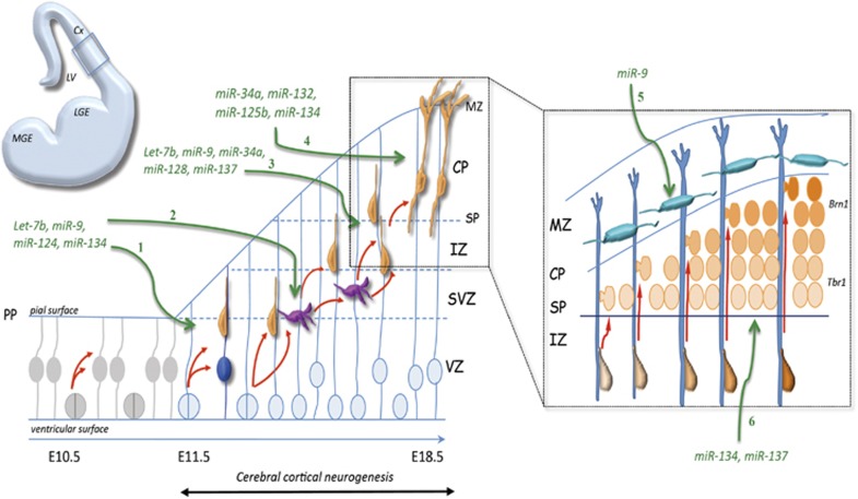
miRNA regulation of cerebral cortical neurogenesis. Scheme representing a part of the cortical wall corresponding to the area boxed in half brain coronal section (upper left). Before onset of neurogenesis (E10.5), symmetrical divisions increase the pool of NEPCs (light gray). Neurogenesis in the Cx starts at E11.5, when some Pax6-positive radial glial cells (blue) divide asymmetrically in the VZ. These cells self-renew (dark blue) and generate neurons in the SVZ either directly (orange) or indirectly through Tbr2-positive basal progenitors (pink). These concurrent steps are regulated by a set of miRNAs: Let-7b, miR-9, miR-124 and miR-134 (1, 2). Basal progenitors can sometime divide asymmetrically to self-renew but also divide symmetrically to give birth to two neurons. Differentiation of projection neurons that migrate through the intermediate zone (IZ) and integrate the CP requires Let-7b, miR-9, miR-34a and miR-137 (3). Terminal differentiation, including dendritogenesis and synapse formation, involves the expression of miR-34a, mir-132, mir-125b and miR-134 (4). The inset (right) shows the inside-out organization of cortical layers. Successive waves of postmitotic neurons are generated (graded orange), and these neurons migrate along radial glial cells (blue) through the IZ to reach the CP. Migration of neurons into the CP is regulated by miR-134 and miR-137 (6). The CP is builded in an inside-out manner and neurons stop their migration under the MZ where Cajal–Retzius cells (CR) (blue turquoise) are located. The generation of CR is controlled by miR-9 (5). MGE, medial ganglionic eminence, LGE, lateral ganglionic eminence, LV, lateral ventricle
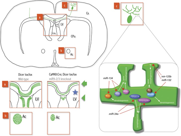
miRNA regulation of axonal pathfinding, dendritogenesis and synaptogenesis. Scheme representing a brain coronal section. Dicer conditional knockout mice (CaMKII:Cre; Dicer:lox/Lox) or miR9-2/3 knockout mice show axonal pathfinding abnormalities (axons are in green). The boxed areas show fasciculation and orientation defects in: (a) the CC (arrow), the F/Hc and the Lsd (arrowhead); (b) the Ac is reduced in mutants. The blue star shows enlarged ventricle in mutant mice. The boxed area in (c) and its zoom-up illustrate a dendrite segment harboring spines. Some miRNAs and their targets regulate dendritogenesis (branching complexity or length, see text), spine morphogenesis (length or width, see text) or synaptogenesis, as indicated on the scheme. Dotted lines refer to target inhibition in hippocampal neurons. LV, lateral ventricle; CPu, caudate-putamen; Lsd, lateral septal dorsal nucleus; 3V, third ventricle
References
-
- Gupta A, Tsai LH, Wynshaw-Boris A. Life is a journey: a genetic look at neocortical development. Nat Rev Genet. 2002;3:342–355. - PubMed
-
- Marin O, Rubenstein JL. Cell migration in the forebrain. Annu Rev Neurosci. 2003;26:441–483. - PubMed
-
- Heng JI. Chariot A, Nguyen L. Molecular layers underlying cytoskeletal remodelling during cortical development. Trends Neurosci. 2010;33:38–47. - PubMed
-
- Kato M, Dobyns WB.Lissencephaly and the molecular basis of neuronal migration Hum Mol Genet 2003. 12 Spec No 1R89–R96. - PubMed
Publication types
MeSH terms
Substances
LinkOut - more resources
Full Text Sources
Molecular Biology Databases
