. 2012 Oct 25:223:77-91.
doi: 10.1016/j.neuroscience.2012.07.043. Epub 2012 Jul 31.

Dorsal root ganglion neurons innervating pelvic organs in the mouse express tyrosine hydroxylase

Affiliations

Dorsal root ganglion neurons innervating pelvic organs in the mouse express tyrosine hydroxylase

P R Brumovsky et al. Neuroscience. .

Abstract

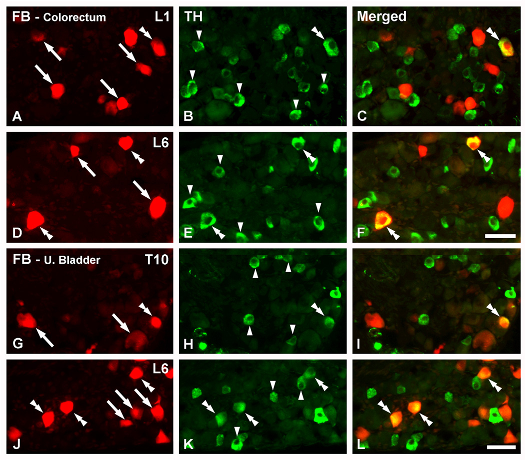
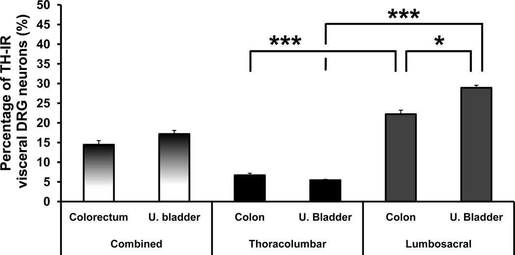
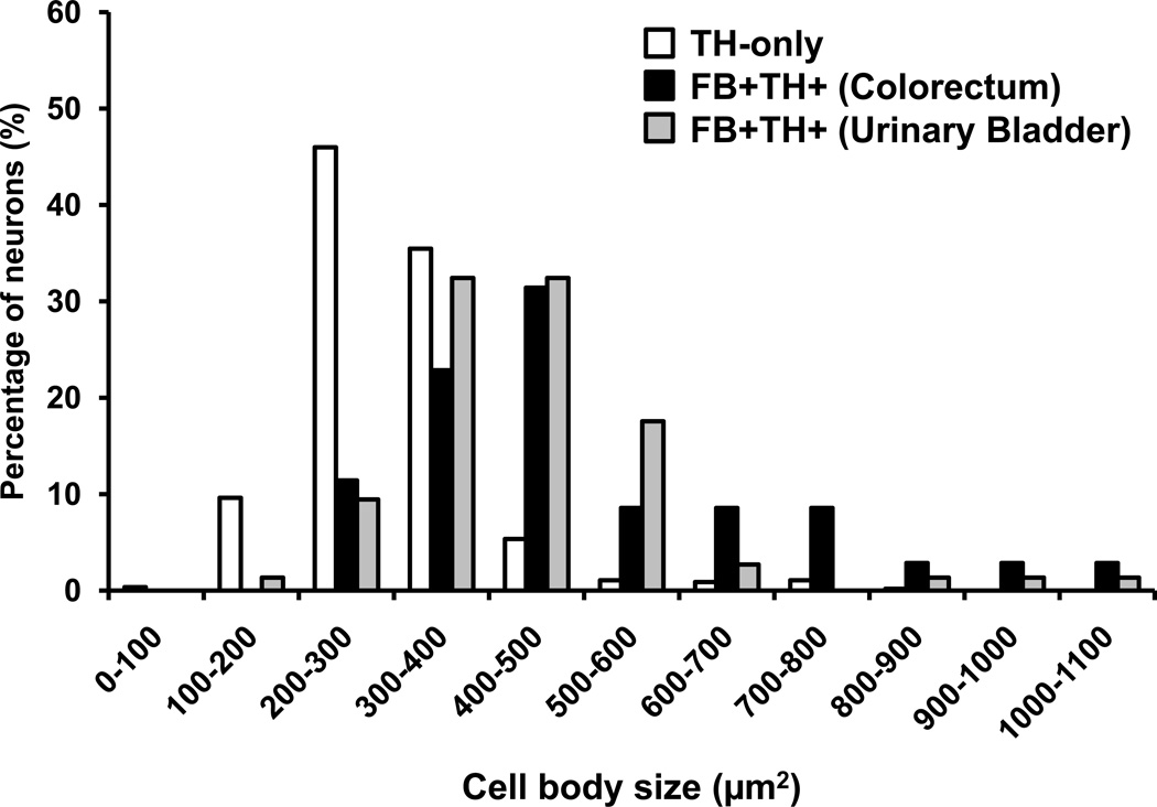
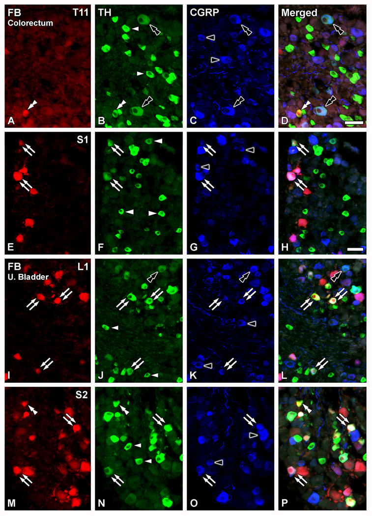
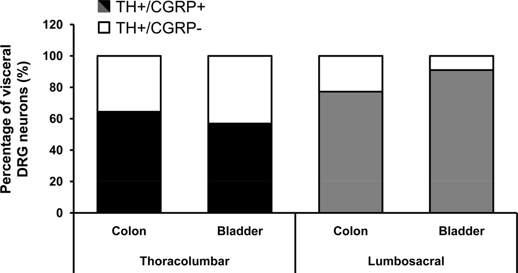
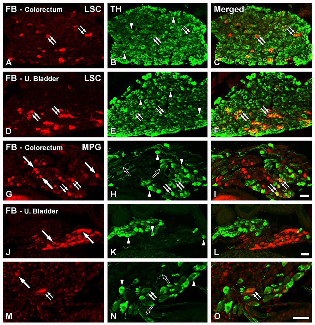
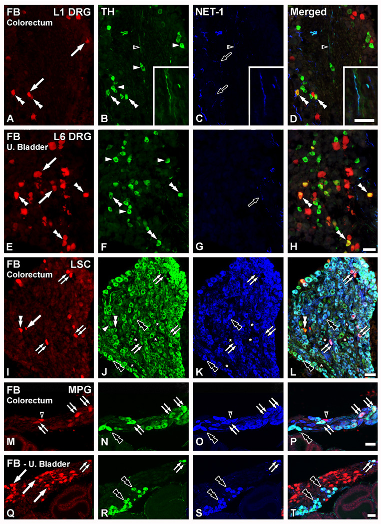
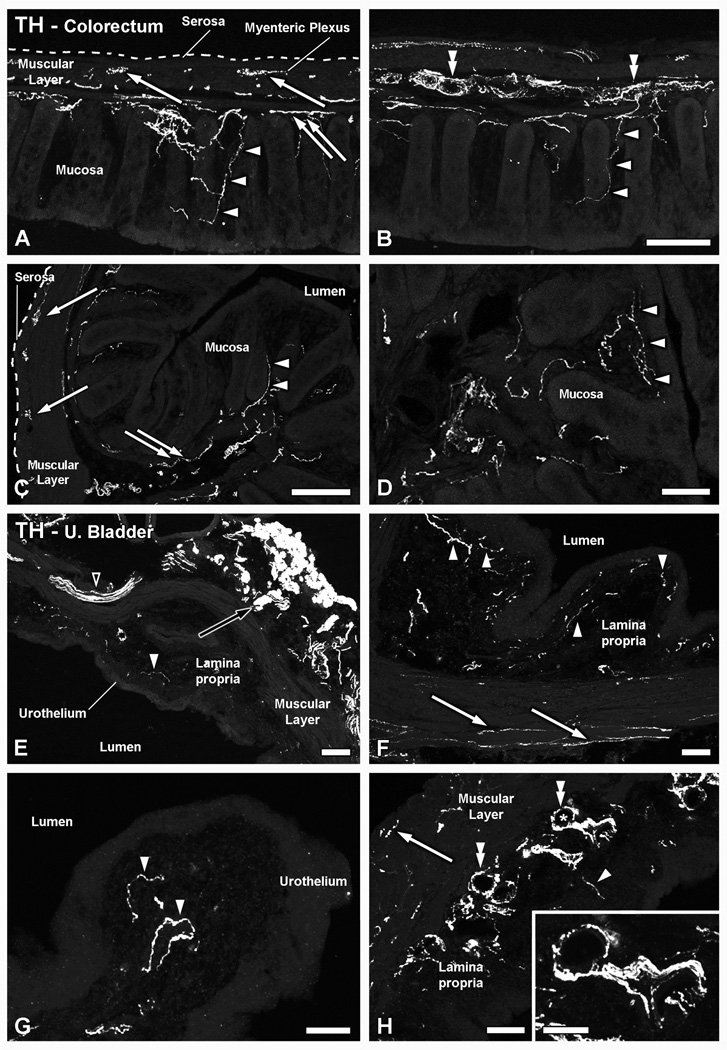
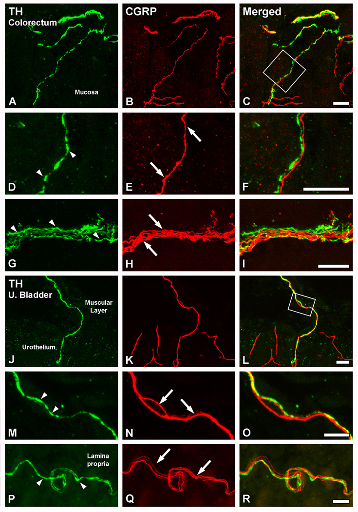
Previous studies in rat and mouse documented that a subpopulation of dorsal root ganglion (DRG) neurons innervating non-visceral tissues express tyrosine hydroxylase (TH). Here we studied whether or not mouse DRG neurons retrogradely traced with Fast Blue (FB) from colorectum or urinary bladder also express immunohistochemically detectable TH. The lumbar sympathetic chain (LSC) and major pelvic ganglion (MPG) were included in the analysis. Previously characterized antibodies against TH, norepinephrine transporter type 1 (NET-1) and calcitonin gene-related peptide (CGRP) were used. On average, ∼14% of colorectal and ∼17% of urinary bladder DRG neurons expressed TH and spanned virtually all neuronal sizes, although more often in the medium-sized to small ranges. Also, they were more abundant in lumbosacral than thoracolumbar DRGs, and often coexpressed CGRP. We also detected several TH-immunoreactive (IR) colorectal and urinary bladder neurons in the LSC and the MPG, more frequently in the former. No NET-1-IR neurons were detected in DRGs, whereas the majority of FB-labeled, TH-IR neurons in the LSC and MPG coexpressed this marker (as did most other TH-IR neurons not labeled from the target organs). TH-IR nerve fibers were detected in all layers of the colorectum and the urinary bladder, with some also reaching the basal mucosal cells. Most TH-IR fibers in these organs lacked CGRP. Taken together, we show: (1) that a previously undescribed population of colorectal and urinary bladder DRG neurons expresses TH, often CGRP but not NET-1, suggesting the absence of a noradrenergic phenotype; and (2) that TH-IR axons/terminals in the colon or urinary bladder, naturally expected to derive from autonomic sources, could also originate from sensory neurons.


Figures

Figure 1
Figure 1
TH is expressed in colorectal and urinary bladder DRG neurons. Optical immunofluorescence photomicrographs of sections of L1 (A–C), L6 (D–F; J–L) or T10 (G–I) DRGs incubated with antiserum to TH. Retrogradely labeled colorectal (A–F) or urinary bladder (G–L) neurons containing FB (A, D, G, J) are shown in red. (C, F, I, L) show merged micrographs. (A–F) A number of colorectal NPs, as evidenced by the presence of FB, express TH in L1 and L6 DRGs (doble arrowheads in A–F). FB+ colorectal NPs lacking the enzyme are also present (arrows in A, D), as well as several TH-only NPs (arrowheads in B, E). (G–L) FB+TH+ urinary bladder NPs are seen in T10 (double arrowheads in G–I) and L6 (double arrrowheads in J–L) DRGs. Also here, FB+ urinary bladder (arrows in G, J) or TH-only (arrowheads in H, K) DRG NPs are detected. Scale bar: 50 µm (F=A–E; L=G–K).
Figure 2
Figure 2
TH is expressed more abundantly in lumbosacral than in thoracolumbar visceral DRG neurons. Percentage of FB+ colorectal or urinary bladder NPs expressing TH, shown as combined (thoracolumbar plus lumbosacral DRGs) or independent values. *, P<0.05; ***, P<0.001.
Figure 3
Figure 3
FB+TH+ DRG neurons tend to be larger than TH-only ones. Graphs showing the size-distribution of TH-only (white bars), FB+TH+ colorectal (black bars) and FB+TH+ urinary bladder (gray bars) DRG neurons in retrogradely traced mice (n = 4). Data are expressed in square µm and include the measurement of 670 DRG NPs (TH-only, 561; FB+TH+ colorectal, 35 neurons; FB+TH+ urinary bladder, 74).
Figure 4
Figure 4
TH-expressing colorectal and urinary bladder DRG neurons colocalize with CGRP. Optical immunofluorescence photomicrographs of sections of T11 (A–D), S1 (E–H), L1 (I–L) or S2 (M–P) DRGs after co-incubation with TH (B, F, J, N) and CGRP antisera (A, E, I, M). Retrogradely labeled colorectal (A–H) or urinary bladder (I–P) neurons containing FB (A, E, I, M) are shown in red. (D, H, L, P) show merged micrographs. (A–P) Several TH-IR NPs are detected (arrowheads in B, F, J, N). Most of the TH-only DRG NPs lack CGRP-LI, with the exception of occasional neurons coexpressing both, the enzyme and the peptide (black double arrowhead in A–D; I–L). Additional non-traced CGRP-IR NPs are also detected (black arrowheads in C, G, K, O). A number of FB+TH+ colorectal (double arrows in E–H) or urinary bladder (double arrows in I–P) NPs are detected, often coexpressing with CGRP. However, some FB+TH+ colorectal (white double arrowhead in A–D) or urinary bladder (white double-arrowhead in M–P) NPs lacking CGRP-LI are also present. Scale bars: 50 µm (D=A-C, M–P; H=E–G; I–L).
Figure 5
Figure 5
A large proportion of TH-expressing colorectal and urinary bladder DRG neurons are peptidergic. Percentages of FB+TH+ colorectal or urinary bladder NPs lacking (white bar segments) or coexpressing (black bar segments) CGRP in thoracolumbar and lumbosacral DRGs.
Figure 6
Figure 6
The main source of autonomic TH fibers in the colorectum and the urinary bladder is the LSC. Optical (A–F; G, J, M) and confocal (H, K, N) immunofluorescence photomicrographs of sections of the LSC (A–F) or MPG (G–O) after incubation with TH (B, E, H, K, N) antiserum. Retrogradely labeled colorectal (A–C; G–I) or urinary bladder (D–F; J–O) neurons containing FB (A, D, G, J, M) are shown in red. (C, F, I, L, O) show merged micrographs. (A–F) A number of FB+TH+ colorectal (A–C) or urinary bladder (D–F) NPs is detected in the LSC (double arrows). In addition, numerous TH-only NPs are also present (arrowheads in B, E). (G–O) In most cases, FB+ colorectal (arrows in G) or urinary bladder (arrows in J, M) NPs lacked TH-LI in the MPG. Likewise, several TH-only NPs could be detected (arrowheads in H, K, N). However, a few TH-IR colorectal (double arrows in G–I) and occasional urinary bladder (double arrows in M–O) NPs are detected. TH-IR fibers were abundant in the MPG (black arrows in H, N). Scale bars: 50 µm (I=A–H; L=J, K; O=M, N).
Figure 7
Figure 7
NET-1 and TH coexpression is observed in the LSC and MPG neurons, but not in DRG neurons. Optical (A–H; I–L; M, Q) and confocal (N, O, R, S) immunofluorescence photomicrographs of sections of L1 (A–D) or L6 (EH) DRGs, the LSC (I–L) or the MPG (M–T) after incubation with TH (B, F, J, N, R) and NET-1 antisera (A, E, I, M, Q). Retrogradely labeled colorectal (A–D; I–P) or urinary bladder (E–H; Q–T) neurons containing FB (A, E, I, M, Q) are shown in red. (D, H, L, P, T) show merged micrographs. (A–H) Neither TH-only (white arrowheads in B, F) nor FB+TH+ (double arrowheads in A–D, E–H) colorectal or urinary bladder NPs in L1 and L6 DRGs coexpressed with NET-1, the latter only present in fiber profiles (black arrows in C, G). Occasional TH-/NET-1-IR fiber profiles were found (black arrowhead in A–D; magnified view in insets B through D). Additional FB+ colorectal (arrows in A) or urinary bladder (arrows in E) NPs lacking TH are also present. (I–L) Virtually all FB+TH+ colorectal (double arrows in I–L), and the vast majority of TH-only LSC NPs (black double arrowheads in I33 L) coexpressed with NET-1. Note, however, a rare FB+TH+ colorectal LSC NP lacking NET-1-LI (white double arrowhead in I–L). Additional TH-only LSC NPs lacking NET-1-LI were also detected (arrowhead in J), as well as a few NPs lacking both markers (asterisks in J–L). A FB+TH+ colorectal neuron lacking both TH and NET-1 is also shown (arrow in I). (M–T) In the MPG, virtually all TH-IR NPs show NET-LI, including colorectal (double arrows in M–P), urinary bladder (double arrows in Q–T) and TH-only NPs (black double arrowheads in M–T). Occasional NET-1-only colorectal NPs could be found (black arrowhead in M–P). Also, several FB+ urinary bladder NPs lacking both TH and NET-1 were often detected (arrows in Q). Scale Bar: 100 µm (H=A–G); 50 µm (L=I–K; P=M–O; T=Q–S); 25 µm (inset in B–D).
Figure 8
Figure 8
Patterns of distribution of TH-IR fibers in the colorectum and the urinary bladder. Confocal immunofluorescence photomicrographs of colorectal (A–D) and urinary bladder (E–H) sections, after incubation with TH antiserum. (A–D) In longitudinal (A, B) as well as coronal (C, D) sections of the colorectum, TH-IR fibers were detected in all layers. Thus, TH-IR fibers could be seen in the muscular layers, in association with the myenteric plexus (arrows in A and C), or in the submucosal layer, either around blood vessels (double arrowheads in B) or in small nerve bundles (double arrows in A, C). A number of TH-IR fibers could also be seen in the colorectal villi, occasionally reaching the basal mucosal cells (arrowheads in A–D). (E–H) In the urinary bladder, thick TH-IR nerve bundles were found penetrating the organ (black arrowhead in E), traveling within the muscular layer (arrows in F, H) or distributed through the lamina propria (white arrowheads in E–H). In the muscular layer, TH-IR nerve profiles appeared sparsely distributed (arrows in F, H). A few of the lamina propria TH-IR fibers were detected in the vicinity of the urothelium (white arrowheads in G). In addition, TH-IR fibers could be observed arranged in thick bundles around blood vessels (asterisk in H, shown at higher magnification in inset) present in the lamina propria (double arrowheads in H). Note the presence of the major pelvic ganglion, strongly immunoreactive for TH (black arrow in E) Scale Bars: 100 µm (B=A; C; E; H); 50 µm (D; F; G; inset in G).
Figure 9
Figure 9
TH and CGRP appear to be present in different populations of nerve fibers innervating the colorectum or the urinary bladder. Confocal immunofluorescence photomicrographs of parasagittal (A–I) and transverse (JR) sections of the colorectum (A–I) or the urinary bladder (J–R), after coincubation with TH (A, D, G, J, M, P) and CGRP (B, E, H, K, N, Q) antisera. (C, F, I, L, O, R) show merged figures. (A–I) In the colorectum, TH- or CGRP-IR fibers were seen in close apposition (A–C). However, closer examination showed that the two markers were present in different nerve populations. This was seen, both in the mucosal layer (inset in C, magnified in D–F), as well as at the level of the myenteric plexus in the muscular layers (G–I). (J–R) In the urinary bladder, and as for the colorectum, TH- (arrowheads in M, P) and CGRP-IR fibers (arrows in N, Q) were found closely juxtaposed with each other (J–L), and virtually always present in different fiber populations. Inset in L is shown at higher magnification in M–L. Scale bars: 20 µm (C=A, B; F=D, E; I= G, H; L=J, K); 10 µm (R=P, Q; O=M, N).

Similar articles

Cited by

References

    1. Adams JC. Biotin amplification of biotin and horseradish peroxidase signals in histochemical stains. J Histochem Cytochem. 1992;40:1457–1463. - PubMed
    1. Biallosterski BT, de Wachter SG, van Koeveringe GA, van Kerrebroeck PE, DE VJ, Mulder MT, Gillespie JI. Changes in bladder innervation in a mouse model of Alzheimer's disease. J. Chem. Neuroanat. 2010;39:204–210. - PubMed
    1. Brierley SM, Carter R, Jones W, III, Xu L, Robinson DR, Hicks GA, Gebhart GF, Blackshaw LA. Differential chemosensory function and receptor expression of splanchnic and pelvic colonic afferents in mice. J Physiol. 2005;567:267–281. - PMC - PubMed
    1. Brierley SM, Jones RC, III, Gebhart GF, Blackshaw LA. Splanchnic and pelvic mechanosensory afferents signal different qualities of colonic stimuli in mice. Gastroenterology. 2004;127:166–178. - PubMed
    1. Brookes SJ, Dinning PG, Gladman MA. Neuroanatomy and physiology of colorectal function and defaecation: from basic science to human clinical studies. Neurogastroenterol. Motil. 2009;21(Suppl 2):9–19. - PubMed

Publication types

Substances