

Influence of exercise training on ischemic brain injury in type 1 diabetic rats
- PMID: 22858624
- PMCID: PMC3774098
- DOI: 10.1152/japplphysiol.00437.2012
Influence of exercise training on ischemic brain injury in type 1 diabetic rats
Abstract
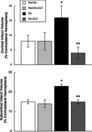
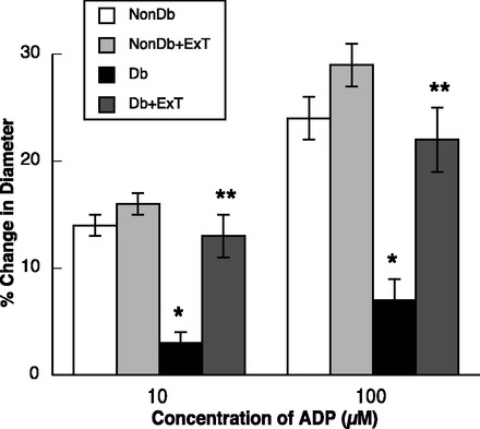
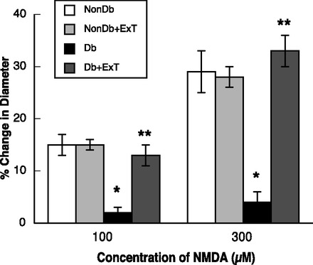
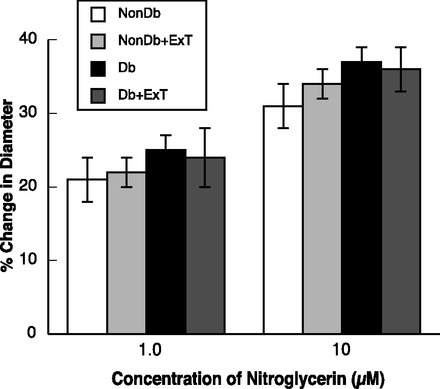
While exercise training (ExT) appears to influence cerebrovascular function during type 1 diabetes (T1D), it is not clear whether this beneficial effect extends to protecting the brain from ischemia-induced brain injury. Thus our goal was to examine whether modest ExT could influence transient focal ischemia-induced brain injury along with nitric oxide synthase (NOS)-dependent dilation of cerebral (pial) arterioles during T1D. Sprague-Dawley rats were divided into four groups: nondiabetic sedentary, nondiabetic ExT, diabetic (streptozotocin; 50 mg/kg ip) sedentary, and diabetic ExT. In the first series of studies, we measured infarct volume in all groups of rats following right MCA occlusion for 2 h, followed by 24 h of reperfusion. In a second series of studies, a craniotomy was performed over the parietal cortex, and we measured responses of pial arterioles to an endothelial NOS (eNOS)-dependent, a neuronal NOS (nNOS)-dependent, and a NOS-independent agonist in all groups of rats. We found that sedentary diabetic rats had significantly larger total, cortical, and subcortical infarct volumes following ischemia-reperfusion than sedentary nondiabetic, nondiabetic ExT, and diabetic ExT rats. Infarct volumes were similar in sedentary nondiabetic, ExT nondiabetic, and ExT diabetic rats. In contrast, ExT did not alter infarct size in nondiabetic compared with sedentary nondiabetic rats. In addition, ExT diabetic rats had impaired eNOS- and nNOS-dependent, but not NOS-independent, vasodilation that was restored by ExT. Thus ExT of T1D rats lessened ischemic brain injury following middle cerebral artery occlusion and restored impaired eNOS- and nNOS-dependent vascular function. Since the incidence of ischemic stroke is increased during T1D, we suggest that our finding are significant in that modest ExT may be a viable preventative therapeutic approach to lessen ischemia-induced brain injury that may occur in T1D subjects.
Figures

References
-
- Ayajiki K, Okamura T, Toda N. Involvement of nitric oxide in endothelium-dependent, phasic relaxation caused by histamine in monkey cerebral arteries. Jpn J Pharmacol 60: 357–362, 1992 - PubMed
-
- Baird TA, Parsons MW, Barber PA, Butcher KS, Desmond PM, Tress BM, Colman PG, Jerums G, Chambers BR, Davis SM. The influence of diabetes mellitus and hyperglycaemia on stroke incidence and outcome. J Clin Neurosci 9: 618–626, 2002 - PubMed
-
- Bravata DM, Kim N, Concato J, Brass LM. Hyperglycaemia in patients with acute ischaemic stroke: how often do we screen for undiagnosed diabetes? QJM 96: 491–497, 2003 - PubMed
-
- Cechetti F, Worm PV, Elsner VR, Bertoldi K, Sanches E, Ben J, Siqueira IR, Netto CA. Forced treadmill exercise prevents oxidative stress and memory deficits following chronic cerebral hypoperfusion in the rat. Neurobiol Learn Mem 97: 90–96, 2012 - PubMed
-
- Chukwuma CS, Tuomilehto J. Diabetes and the risk of stroke. J Diabetes Complications 7: 250–262, 1993 - PubMed
Publication types
MeSH terms
Substances
Grants and funding
LinkOut - more resources
Full Text Sources
Medical