. 2012 Aug;9(8):1066-75.
doi: 10.4161/rna.21083. Epub 2012 Aug 1.

Distinct AGO1 and AGO2 associated miRNA profiles in human cells and blood plasma

Affiliations

Distinct AGO1 and AGO2 associated miRNA profiles in human cells and blood plasma

Andrey Turchinovich et al. RNA Biol. 2012 Aug.

Abstract

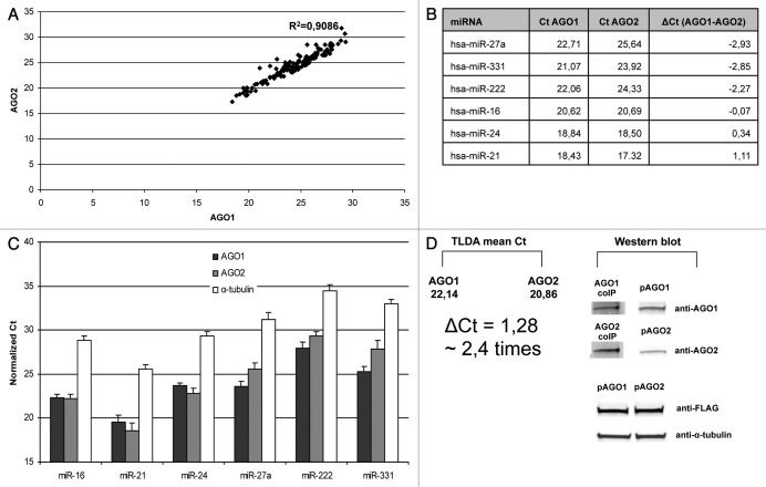
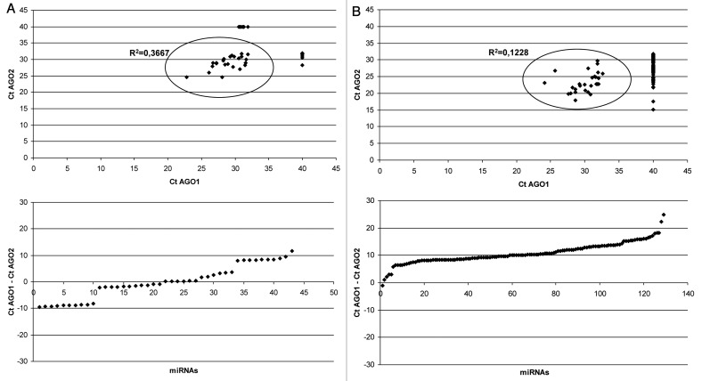
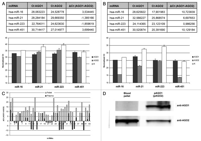
Studies of miRNA association with Argonaute (AGO) proteins in mammalian cells have indicated lack of bias toward particular AGO. However, to our knowledge, the use of quantitative methods for studying miRNA association with different AGOs has not been reported so far. In this work we compared the total miRNA content in AGO1 and AGO2 immunoprecipitates obtained from MCF7 adenocarcinoma cells using TaqMan Low Density miRNA Arrays and successfully verified selected miRNAs with qPCR. For most of the miRNA species AGO1 and AGO2 profiles were well correlated, however, some miRNAs demonstrated consistent biases toward one of the Argonautes. Furthermore, miRNAs which were predominantly AGO2-associated derived mostly from sense strands of the corresponding pre-miRNAs while the majority of AGO1 biased miRNAs originated from antisense strands of the pre-miRNAs. Additionally, we show that circulating miRNA in human blood plasma can be immunoprecipitated with both AGO1 and AGO2 antibody. However, unlike in cell lysates, AGO1 and AGO2 associated miRNA profiles in plasma did not correlate, indicating that many cell types contribute to circulating miRNA (given that expression of AGO proteins is tissue specific). Furthermore, AGO-specific miRNA profiles in blood cells differed significantly from miRNAs profiles in plasma indicating that most circulating miRNAs are likely to derive from non-blood cells. Since circulating miRNAs hold great promise as biomarkers for numerous cancers and other diseases, we hypothesize that AGO-specific miRNA profiles might add an additional dimension to circulating miRNA-based diagnostics.


Figures

None
Figure 1. Association of miRNAs with endogenous AGO1 and AGO2 proteins in MCF7 cells. (A) Correlation plot showing Ct values of miRNA signals detected in anti-AGO1 and anti-AGO2 pellets using TaqMan Low Density miRNA Arrays (TLDA). Note, the high correlation coefficient indicate low bias in binding toward AGO1 and AGO2 proteins for most miRNAs. (B) Ct values of the miRNA species on TLDA which were selected for verification with individual qPCR assays. (C) Individual TaqMan miRNA Assays of miR-16, miR-21, miR-24, miR-27a, miR-331 and miR-222 in anti-AGO1, anti-AGO2 and anti-α-tubulin (control) immunoprecipitates from MCF7 cells. Spiked in cel-miR-39 was used as control for isolation efficiency in all samples. Data are presented as Ct values normalized on both cel-miR-39 and the mean Ct of [miR-16, miR-21, miR-24, miR-27a, miR-331 and miR-222] within each sample (quantile normalization). Each bar represents mean + SD of three independent experiments. (D) Estimation of average miRNA association with each AGO protein. The means of Ct values of miRNAs in either anti-AGO1 or anti-AGO2 samples detected on the array (left) were compared with the relative content of AGO1 and AGO2 proteins detected by western blot (right) in the same immunopellets. To account for the difference in anti-AGO1 and anti-AGO2 antibody efficiency on western blot, equal amounts of recombinant FLAG-AGO1 (pAGO1) and FLAG-AGO2 (pAGO2) proteins were applied on the SDS gel. Note, the content of AGO1 protein in the anti-AGO1 coIP pellet was lower as compared with AGO2 protein in anti-AGO2 coIP pellet (~2,5 times as analyzed by densitometry). Accordingly, the observed difference in the mean Ct values between AGO1 and AGO2 coIP pellets corresponded to approximately 2,5 times difference in the total miRNA content.
None
Figure 2. TaqMan miRNA Low Density Arrays performed on total RNA extracted from anti-AGO1 and anti-AGO2 immunoprecipitates of blood plasma (A) and whole blood pellet (B). Blood plasma (80 μL) was combined with 20 μg the antibody. Blood pellet lysate (5 μL) combined with 3 μg of the antibody. MiRNAs which were undetected on the array (Ct > 32) were assigned Ct values of 40. Note, (1) some miRNAs were presented exclusively in the coIP pellets of particular AGO protein; (2) unlike in blood plasma, in blood pellet overwhelming majority of miRNAs were AGO2 associated.
None
Figure 3. (A) miRNA species in the blood plasma which showed significant bias toward either AGO1 (miR-21, miR-223) or AGO2 (miR-16, miR-451) proteins on TaqMan Low Density miRNA Array (above) and their verification with individual miRNA qPCR assays (below) in three independent experiments; (B) The same miRNA species demonstrated drastically different AGO1/AGO2 distribution in whole blood pellet: TLDA (above), individual qPCR assays (below). Data presented as Ct values normalized on cel-miR-39 and miR-16. Each bar represents mean + SD of three independent experiments (samples from different individuals). (C) The ΔCt (AGO1-AGO2) graph of miRNAs detected in both blood plasma and blood pellet. Note, for most of the miRNA there is no correlation of ΔCt (AGO1-AGO2) between plasma and pellet; (D) western blot analysis of AGO1 and AGO2 proteins in anti-AGO1 and anti-AGO2 coIP samples respectively. Note, unlike AGO2 protein, the amount of AGO1 in the immuprecipitate was below the detection limit.

References

    1. Bartel DP. MicroRNAs: genomics, biogenesis, mechanism, and function. Cell. 2004;116:281–97. doi: 10.1016/S0092-8674(04)00045-5. - DOI - PubMed
    1. Lee Y, Ahn C, Han J, Choi H, Kim J, Yim J, et al. The nuclear RNase III Drosha initiates microRNA processing. Nature. 2003;425:415–9. doi: 10.1038/nature01957. - DOI - PubMed
    1. Han J, Lee Y, Yeom KH, Kim YK, Jin H, Kim VN. The Drosha-DGCR8 complex in primary microRNA processing. Genes Dev. 2004;18:3016–27. doi: 10.1101/gad.1262504. - DOI - PMC - PubMed
    1. Hutvágner G, McLachlan J, Pasquinelli AE, Bálint E, Tuschl T, Zamore PD. A cellular function for the RNA-interference enzyme Dicer in the maturation of the let-7 small temporal RNA. Science. 2001;293:834–8. doi: 10.1126/science.1062961. - DOI - PubMed
    1. Han J, Lee Y, Yeom KH, Nam JW, Heo I, Rhee JK, et al. Molecular basis for the recognition of primary microRNAs by the Drosha-DGCR8 complex. Cell. 2006;125:887–901. doi: 10.1016/j.cell.2006.03.043. - DOI - PubMed

Publication types

LinkOut - more resources