. 2012 Sep 28;287(40):33836-46.
doi: 10.1074/jbc.M112.394841. Epub 2012 Aug 2.

Structural basis of Rev1-mediated assembly of a quaternary vertebrate translesion polymerase complex consisting of Rev1, heterodimeric polymerase (Pol) ζ, and Pol κ

Affiliations

Structural basis of Rev1-mediated assembly of a quaternary vertebrate translesion polymerase complex consisting of Rev1, heterodimeric polymerase (Pol) ζ, and Pol κ

Jessica Wojtaszek et al. J Biol Chem. .

Abstract

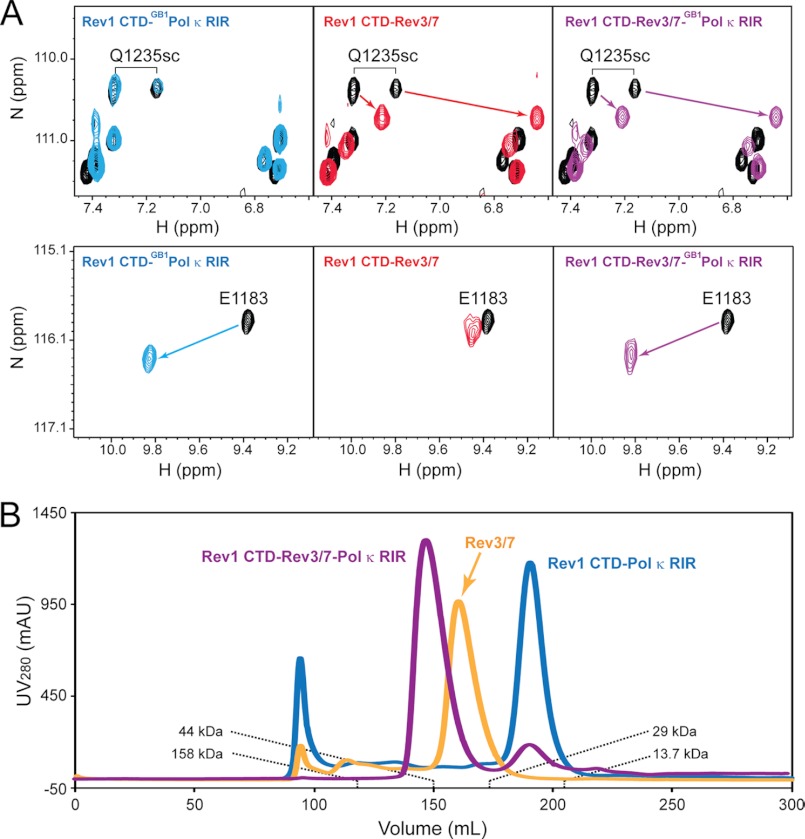
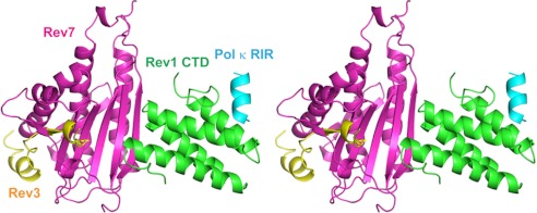
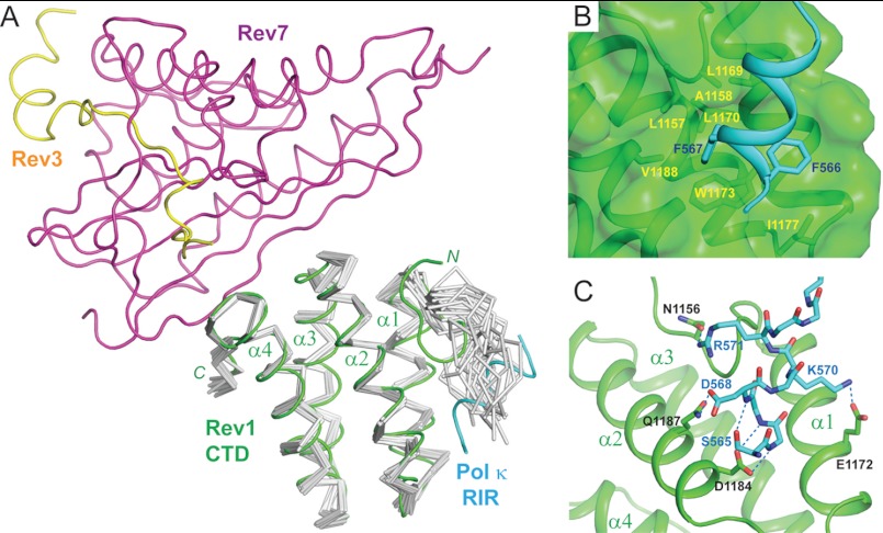
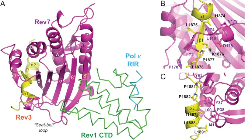
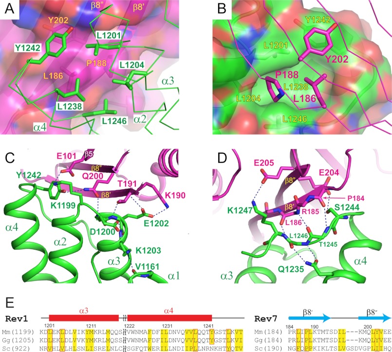
DNA synthesis across lesions during genomic replication requires concerted actions of specialized DNA polymerases in a potentially mutagenic process known as translesion synthesis. Current models suggest that translesion synthesis in mammalian cells is achieved in two sequential steps, with a Y-family DNA polymerase (κ, η, ι, or Rev1) inserting a nucleotide opposite the lesion and with the heterodimeric B-family polymerase ζ, consisting of the catalytic Rev3 subunit and the accessory Rev7 subunit, replacing the insertion polymerase to carry out primer extension past the lesion. Effective translesion synthesis in vertebrates requires the scaffolding function of the C-terminal domain (CTD) of Rev1 that interacts with the Rev1-interacting region of polymerases κ, η, and ι and with the Rev7 subunit of polymerase ζ. We report the purification and structure determination of a quaternary translesion polymerase complex consisting of the Rev1 CTD, the heterodimeric Pol ζ complex, and the Pol κ Rev1-interacting region. Yeast two-hybrid assays were employed to identify important interface residues of the translesion polymerase complex. The structural elucidation of such a quaternary translesion polymerase complex encompassing both insertion and extension polymerases bridged by the Rev1 CTD provides the first molecular explanation of the essential scaffolding function of Rev1 and highlights the Rev1 CTD as a promising target for developing novel cancer therapeutics to suppress translesion synthesis. Our studies support the notion that vertebrate insertion and extension polymerases could structurally cooperate within a megatranslesion polymerase complex (translesionsome) nucleated by Rev1 to achieve efficient lesion bypass without incurring an additional switching mechanism.


Figures

FIGURE 1.
FIGURE 1.
Formation of a quaternary complex consisting of the Rev1 CTD, Rev3/7, and Pol κ RIR. A, 1H-15N HSQC spectra of the free Rev1 CTD (black) and the Rev1 CTD in the presence of an equal molar ratio of either the GB1-Pol κ RIR fusion protein (blue), Rev3/7 (red), or both (purple). B, FPLC traces of the Rev1 CTD-Rev3/7-Pol κ RIR complex (purple), the Rev3/7 complex (light orange), and the Rev1 CTD-Pol κ RIR complex (blue) separated using a HiPrep 26/60 Sephacryl S-200 HR column (GE Healthcare). The elution volumes of known protein standards are labeled.
FIGURE 2.
FIGURE 2.
Structure of the quaternary Rev1 CTD-Rev3/7-Pol κ RIR complex. Ribbon diagrams of the complex are shown in stereo view and are colored with the Rev1 CTD in green, Rev3 in yellow, Rev7 in purple, and the Pol κ RIR in cyan.
FIGURE 3.
FIGURE 3.
The Rev1 CTD-Pol κ RIR interface. A, structure of the Rev1 CTD-Rev3/7-Pol κ RIR complex superimposed with the NMR ensemble of the free Rev1 CTD (PDB ID: 2LSG). Components of the quaternary complex are colored, with the Rev1 CTD in green, Pol κ RIR in cyan, Rev7 in purple, and Rev3 in yellow. The NMR ensemble of the free Rev1 CTD is shown in Cα traces and colored gray. B, hydrophobic interactions between the Rev1 CTD and Pol κ RIR. C, hydrophilic interactions between the Rev1 CTD and Pol κ RIR.
FIGURE 4.
FIGURE 4.
The Rev3/7 interface. A, overall structure of the Rev3/7 complex in the quaternary Rev1 CTD-Rev3/7-Pol κ RIR complex. Rev3 and Rev7 are shown in a ribbon diagram, and the Rev1 CTD and Pol κ RIR are shown in Cα traces. B, Rev3-Rev7 binding interface encompassing the N-terminal α1 helix and the β1 strand of Rev3. C, Rev3-Rev7 interface encompassing the C-terminal α2 helix and the loop connecting the β1 strand and the α2 helix of Rev3.
FIGURE 5.
FIGURE 5.
The Rev1 CTD-Rev7 interface. A, hydrophobic interactions between the Rev1 CTD and Rev7. Rev7 residues are labeled in yellow, and Rev1 residues are labeled in green. B, opposite view of A, illustrating the central hydrophobic pocket of the Rev1 CTD that accommodates Pro-188 and Leu-186 of Rev7. Rev7 residues are labeled in purple, and Rev1 residues are labeled in yellow. C and D, hydrophilic interactions between the Rev1 CTD and Rev7, with C showing interactions centered at the α2-α3 loop of the Rev1 CTD and D showing interactions centered at the C-terminal tail of the Rev1 CTD. E, sequence alignment of the Rev1 CTD and Rev7 from mouse (Mus musculus; Mm), chicken (Gallus gallus; Gg), and yeast (S. cerevisiae; Sc). Conserved hydrophobic residues are colored yellow. Hydrophobic residues important for the Rev1-Rev7 interaction are boxed in red.
FIGURE 6.
FIGURE 6.
Rev1 is critical for DNA damage tolerance in vertebrate cells. A, wild-type chicken DT40 cells (DT40) and Rev1 deletion chicken DT40 cells (Rev1) harboring full-length wild-type Rev1 (Rev1+WT) or the K1199E mutation (Rev1+K1199E) were treated with the indicated doses of cisplatin (cis-diammineplatinum(II) dichloride), and cell viability was measured 72 h later. B, immunoblot analysis of lysates prepared from Rev1 deletion chicken DT40 cells (−), cells expressing full-length GFP-tagged wild-type Rev1 (Wild-type), and the K1199E mutation (K1199E) probed with an anti-GFP antibody. The same blot was probed with an anti-β-actin antibody as a loading control. Error bars represent standard deviation from three independent experiments.

References

    1. Friedberg E. C., Walker G. C., Siede W., Wood R. D., Schultz R. A., Ellenberger T. (eds) (2005) DNA Repair and Mutagenesis, American Society for Microbiology, Washington, D. C
    1. Waters L. S., Minesinger B. K., Wiltrout M. E., D'Souza S., Woodruff R. V., Walker G. C. (2009) Eukaryotic translesion polymerases and their roles and regulation in DNA damage tolerance. Microbiol. Mol. Biol. Rev. 73, 134–154 - PMC - PubMed
    1. Sale J. E., Lehmann A. R., Woodgate R. (2012) Y-family DNA polymerases and their role in tolerance of cellular DNA damage. Nat. Rev. Mol. Cell Biol. 13, 141–152 - PMC - PubMed
    1. Lemontt J. F. (1971) Mutants of yeast defective in mutation induced by ultraviolet light. Genetics 68, 21–33 - PMC - PubMed
    1. Lawrence C. W., Das G., Christensen R. B. (1985) REV7, a new gene concerned with UV mutagenesis in yeast. Mol. Gen. Genet. 200, 80–85 - PubMed

Publication types

MeSH terms

Associated data