

Intrafibrillar silicification of collagen scaffolds for sustained release of stem cell homing chemokine in hard tissue regeneration
- PMID: 22859369
- PMCID: PMC3475249
- DOI: 10.1096/fj.12-210211
Intrafibrillar silicification of collagen scaffolds for sustained release of stem cell homing chemokine in hard tissue regeneration
Abstract
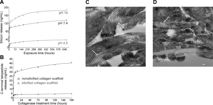
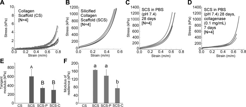
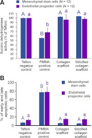
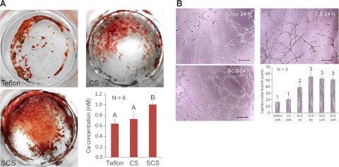
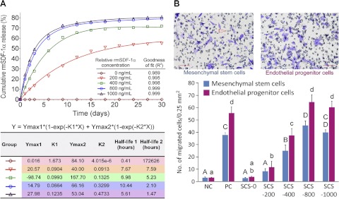
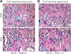
Traditional bone regeneration strategies relied on supplementation of biomaterials constructs with stem or progenitor cells or growth factors. By contrast, cell homing strategies employ chemokines to mobilize stem or progenitor cells from host bone marrow and tissue niches to injured sites. Although silica-based biomaterials exhibit osteogenic and angiogenic potentials, they lack cell homing capability. Stromal cell-derived factor-1 (SDF-1) plays a pivotal role in mobilization and homing of stem cells to injured tissues. In this work, we demonstrated that 3-dimensional collagen scaffolds infiltrated with intrafibrillar silica are biodegradable and highly biocompatible. They exhibit improved compressive stress-strain responses and toughness over nonsilicified collagen scaffolds. They are osteoconductive and up-regulate expressions of osteogenesis- and angiogenesis-related genes more significantly than nonsilicified collagen scaffolds. In addition, these scaffolds reversibly bind SDF-1α for sustained release of this chemokine, which exhibits in vitro cell homing characteristics. When implanted subcutaneously in an in vivo mouse model, SDF-1α-loaded silicified collagen scaffolds stimulate the formation of ectopic bone and blood capillaries within the scaffold and abrogate the need for cell seeding or supplementation of osteogenic and angiogenic growth factors. Intrafibrillar-silicified collagen scaffolds with sustained SDF-1α release represent a less costly and complex alternative to contemporary cell seeding approaches and provide new therapeutic options for in situ hard tissue regeneration.
Figures

Similar articles
-
Intrafibrillar-silicified collagen scaffolds enhance the osteogenic capacity of human dental pulp stem cells.J Dent. 2014 Jul;42(7):839-49. doi: 10.1016/j.jdent.2014.03.016. Epub 2014 Apr 3. J Dent. 2014. PMID: 24705068
-
Intrafibrillar silicified collagen scaffold modulates monocyte to promote cell homing, angiogenesis and bone regeneration.Biomaterials. 2017 Jan;113:203-216. doi: 10.1016/j.biomaterials.2016.10.050. Epub 2016 Oct 31. Biomaterials. 2017. PMID: 27821306
-
Mineralised Collagen Scaffolds Loaded with Stromal Cell-derived Factor-1 Improve Mandibular Bone Regeneration.Chin J Dent Res. 2014;17(1):23-9. Chin J Dent Res. 2014. PMID: 25028686
-
Strategies to develop endogenous stem cell-recruiting bioactive materials for tissue repair and regeneration.Adv Drug Deliv Rev. 2017 Oct 1;120:50-70. doi: 10.1016/j.addr.2017.07.011. Epub 2017 Jul 19. Adv Drug Deliv Rev. 2017. PMID: 28734899 Free PMC article. Review.
-
In situ tissue regeneration through host stem cell recruitment.Exp Mol Med. 2013 Nov 15;45(11):e57. doi: 10.1038/emm.2013.118. Exp Mol Med. 2013. PMID: 24232256 Free PMC article. Review.
Cited by
-
Multiphase intrafibrillar mineralization of collagen.Angew Chem Int Ed Engl. 2013 May 27;52(22):5762-6. doi: 10.1002/anie.201210259. Epub 2013 Apr 18. Angew Chem Int Ed Engl. 2013. PMID: 23606345 Free PMC article. No abstract available.
-
Biphasic silica/apatite co-mineralized collagen scaffolds stimulate osteogenesis and inhibit RANKL-mediated osteoclastogenesis.Acta Biomater. 2015 Jun;19:23-32. doi: 10.1016/j.actbio.2015.03.012. Epub 2015 Mar 16. Acta Biomater. 2015. PMID: 25792280 Free PMC article.
-
Scaffold design for bone regeneration.J Nanosci Nanotechnol. 2014 Jan;14(1):15-56. doi: 10.1166/jnn.2014.9127. J Nanosci Nanotechnol. 2014. PMID: 24730250 Free PMC article. Review.
-
Effect of the nano/microscale structure of biomaterial scaffolds on bone regeneration.Int J Oral Sci. 2020 Feb 6;12(1):6. doi: 10.1038/s41368-020-0073-y. Int J Oral Sci. 2020. PMID: 32024822 Free PMC article. Review.
-
Decellularized Hydrogels in Bone Tissue Engineering: A Topical Review.Int J Med Sci. 2018 Mar 8;15(5):492-497. doi: 10.7150/ijms.22789. eCollection 2018. Int J Med Sci. 2018. PMID: 29559838 Free PMC article. Review.
References
-
- Dhandayuthapani B., Yoshida Y., Maekawa T., Kumar D. S. (2011) Polymeric scaffolds in tissue engineering application: a review. Int. J. Polym. Sci. 2011, 290602
-
- Teo A. K., Vallier L. (2010) Emerging use of stem cells in regenerative medicine. Biochem. J. 428, 11–23 - PubMed
-
- Chen F. M., Wu L. A., Zhang M., Zhang R., Sun H. H. (2011) Homing of endogenous stem/progenitor cells for in situ tissue regeneration: Promises, strategies, and translational perspectives. Biomaterials 32, 3189–3209 - PubMed
Publication types
MeSH terms
Substances
Grants and funding
LinkOut - more resources
Full Text Sources
