. 2012 Oct 15;186(8):780-9.
doi: 10.1164/rccm.201203-0411OC. Epub 2012 Aug 2.

Dysregulated renin-angiotensin-aldosterone system contributes to pulmonary arterial hypertension

Affiliations
Free PMC article

Dysregulated renin-angiotensin-aldosterone system contributes to pulmonary arterial hypertension

Frances S de Man et al. Am J Respir Crit Care Med. .
Free PMC article

Abstract

Rationale: Patients with idiopathic pulmonary arterial hypertension (iPAH) often have a low cardiac output. To compensate, neurohormonal systems such as the renin-angiotensin-aldosterone system (RAAS) and the sympathetic nervous system are up-regulated, but this may have long-term negative effects on the progression of iPAH.

Objectives: Assess systemic and pulmonary RAAS activity in patients with iPAH and determine the efficacy of chronic RAAS inhibition in experimental PAH.

Methods: We collected 79 blood samples from 58 patients with iPAH in the VU University Medical Center Amsterdam (between 2004 and 2010) to determine systemic RAAS activity.

Measurements and main results: We observed increased levels of renin, angiotensin (Ang)I, and AngII, which were associated with disease progression (P < 0.05) and mortality (P < 0.05). To determine pulmonary RAAS activity, lung specimens were obtained from patients with iPAH (during lung transplantation, n = 13) and control subjects (during lobectomy or pneumonectomy for cancer, n = 14). Local RAAS activity in pulmonary arteries of patients with iPAH was increased, demonstrated by elevated angiotensin-converting enzyme activity in pulmonary endothelial cells and increased AngII type 1 (AT(1)) receptor expression and signaling. In addition, local RAAS up-regulation was associated with increased pulmonary artery smooth muscle cell proliferation via enhanced AT(1) receptor signaling in patients with iPAH compared with control subjects. Finally, to determine the therapeutic potential of RAAS activity, we assessed the chronic effects of an AT(1) receptor antagonist (losartan) in the monocrotaline PAH rat model (60 mg/kg). Losartan delayed disease progression, decreased right ventricular afterload and pulmonary vascular remodeling, and restored right ventricular-arterial coupling in rats with PAH.

Conclusions: Systemic and pulmonary RAAS activities are increased in patients with iPAH and are associated with increased pulmonary vascular remodeling. Chronic inhibition of RAAS by losartan is beneficial in experimental PAH.


Figures

Figure 1
Figure 1. Serum RAAS in iPAH-patients
Serum levels of renin, AngI and AngII were increased in the majority of patients in comparison to upper limit of normal (dotted line; A–C). Follow-up measurements in a subgroup of patients (n=21) revealed that increased serum levels of renin, AngI and AngII were associated with progressive iPAH (D–F). Data presented as mean ±SEM, n=58 iPAH-patients. Dotted line represents the upper limit of reference. Light blue bars represent baseline, dark blue bars represent follow up. iPAH, idiopathic pulmonary arterial hypertension.
Figure 2
Figure 2. Pulmonary endothelial cells of iPAH-patients produce more angiotensin II
Exposure of pulmonary endothelial cells to AngI revealed significant higher production of AngII in pulmonary endothelial cells of iPAH-patients than controls. Co-incubation with ACE-inhibitor enalapril totally abolished this effect, indicating that ACE-activity in pulmonary endothelial cells of iPAH-patients is increased. ** p<0.01, ***p<0.001 vs. values of unstimulated cells (Base). Data presented as mean ±SEM, n=3 per group. CON, control; iPAH, idiopathic pulmonary arterial hypertension; Base, unstimulated condition; AngI, angiotensin I; Enalapril, ACE-inhibitor.
Figure 3
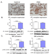
Figure 3. Angiotensin II type 1 receptor expression and signaling is increased in pulmonary arteries of iPAH-patients
Typical examples of histological sections of lung specimens are shown of a control and iPAH-patient (A), stained for angiotensin II type 1 receptor (AT1-receptor). Western blot analyses revealed significant upregulation of AT1-receptor expression in pulmonary arteries of iPAH-patients in comparison to control; no changes in AT2-receptor expression were observed (B). In addition, tyrosine kinase SRC-activity and ERK-activity (downstream targets of AT1-receptor) were significantly increased, suggesting increased signaling activity of the AT1-receptor (C). Data presented as mean ±SEM, n=5 per group. AT1-receptor, angiotensin II type 1 receptor; p-SRC, expression phosphorylated form of tyrosine kinase SRC; t-SRC, total protein expression of tyrosine kinase SRC; p-ERK, expression phosphorylated form of extracellular regulated kinase; t-ERK, total protein expression of extracellular regulated kinase; P, idiopathic pulmonary arterial hypertension; C, control; M, marker.
Figure 4
Figure 4. Angiotensin II incubation induces selective proliferation of the pulmonary artery smooth muscle cells of iPAH-patients via AT1-receptor signaling
Exposure of pulmonary artery smooth muscle cells (PA-SMC) to angiotensin II induced significantly more PA-SMC proliferation in iPAH-patients in comparison to controls. Co-incubation with an AT1-receptor antagonist (losartan) abolished this effect completely. This indicates that angiotensin II exerts its proliferative effect in PA-SMC of iPAH-patients via AT1-receptor signaling. ** p<0.01; *** p<0.001 vs. values of unstimulated cells (Base). Data presented as mean ±SEM, n=4 per group. iPAH, idiopathic pulmonary arterial hypertension; PA-SMC, pulmonary artery smooth muscle cells; Base, unstimulated condition; FCS, fetal calf serum; AngII, angiotensin II; losartan, AT1-receptor antagonist
Figure 5
Figure 5. Losartan dose finding in control and PAH rats
Maximal tolerated dosage (<10% reduction in aortic pressure; dotted line) of losartan was tested by 48-hours telemetry registration. Four different dosages of losartan were tested in control rats (total n=9): 5 mg/kg (n=2); 10 mg/kg (n=3); 20 mg/kg (n=2); 40 mg/kg (n=2). After MCT-injection, dose finding was repeated with three different dosages: 5 mg/kg (n=3); 10 mg/kg (n=3); 20 mg/kg (n=3). A dose of 40 mg/kg was not tested in PAH-rats due to >10% reduction in aortic pressure in control rats. All dosages tested in PAH-rats did not induce >10% reduction in aortic pressure. A dose of 20 mg/kg was therefore used to test the chronic effects of losartan in PAH-rats. Data presented as mean ±SEM, n=2/3 per group. PAH, pulmonary arterial hypertension; ΔP aorta, change in aortic pressure.
Figure 6
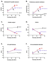
Figure 6. Losartan significantly delayed disease progression in PAH-treated rats
Losartan treatment delayed the progression of pulmonary vascular remodeling (A, B), and delayed RV dilation (F). No changes were observed in cardiac function (C,D) or RV wall thickness (E). Data presented as mean ±SEM, n=9 per group. ## p<0.01, ### p<0.001 PAH/PAH+losartan vs. control, * p<0.05, ** p<0.01 PAH vs. PAH+losartan. RV, right ventricle; TAPSE, tricuspid annular plane systolic excursion; MCT, monocrotaline; Con, control; PAH, pulmonary arterial hypertension.
Figure 7
Figure 7. Losartan significantly reduced RV afterload, restore ventricular-arterial coupling and improved RV diastolic function
Typical examples of pressure-volume relation are shown for control, PAH and PAH+losartan (A–C). Loasartan reduced RV arterial elastance (D) without affecting RV contractility (E). In addition, RV diastolic function improved significantly after losartan treatment illustrated by reduced RV end-diastolic elastance (F). For measurements of RV dilatation, see Figure 6. Data presented as mean ±SEM, n=9 per group. Con, control; PAH, pulmonary arterial hypertension.
Figure 8
Figure 8. Losartan treatment reduced muscularization of the pulmonary arterioles without changing RV hypertrophy
Representative images of histological sections of lung specimens are shown of control, PAH and PAH+losartan rats (A), stained for WGA (glycocalyx, red). Losartan significantly reduced pulmonary artery wall thickness in PAH rats (B). RV cross sectional area was equally increased in both PAH and PAH+losartan rats (C). Data presented as mean ±SEM, n=9 per group. PA, pulmonary artery; CSA, cross sectional area; CON, control; PAH, pulmonary arterial hypertension.

Comment in

References

    1. Humbert M, Sitbon O, Chaouat A, Bertocchi M, Habib G, Gressin V, Yaici A, Weitzenblum E, Cordier JF, Chabot F, Dromer C, Pison C, Reynaud-Gaubert M, Haloun A, Laurent M, Hachulla E, Cottin V, Degano B, Jais X, Montani D, Souza R, Simonneau G. Survival in patients with idiopathic, familial, and anorexigen-associated pulmonary arterial hypertension in the modern management era. Circulation. 2010;122:156–163. - PubMed
    1. Handoko ML, de Man FS, Allaart CP, Paulus WJ, Westerhof N, Vonk-Noordegraaf A. Perspectives on novel therapeutic strategies for right heart failure in pulmonary arterial hypertension: Lessons from the left heart. Eur Respir Rev. 2010;19:72–82. - PMC - PubMed
    1. Eichhorn EJ, Bristow MR. Medical therapy can improve the biological properties of the chronically failing heart. A new era in the treatment of heart failure. Circulation. 1996;94:2285–2296. - PubMed
    1. Ciarka A, Doan V, Velez-Roa S, Naeije R, van de Borne P. Prognostic significance of sympathetic nervous system activation in pulmonary arterial hypertension. Am J Respir Crit Care Med. 2010;181:1269–1275. - PubMed
    1. de Man FS, Handoko ML, van Ballegoij JJ, Schalij I, Bogaards SJ, Postmus PE, van dV, Westerhof N, Paulus WJ, Vonk-Noordegraaf A. Bisoprolol delays progression towards right heart failure in experimental pulmonary hypertension. Circ Heart Fail. 2012;5:97–105. - PubMed

Publication types

MeSH terms