. 2012 Oct 11;120(15):3048-57.
doi: 10.1182/blood-2012-06-435297. Epub 2012 Aug 2.

STAT3 mutations unify the pathogenesis of chronic lymphoproliferative disorders of NK cells and T-cell large granular lymphocyte leukemia

Affiliations

STAT3 mutations unify the pathogenesis of chronic lymphoproliferative disorders of NK cells and T-cell large granular lymphocyte leukemia

Andres Jerez et al. Blood. .

Abstract

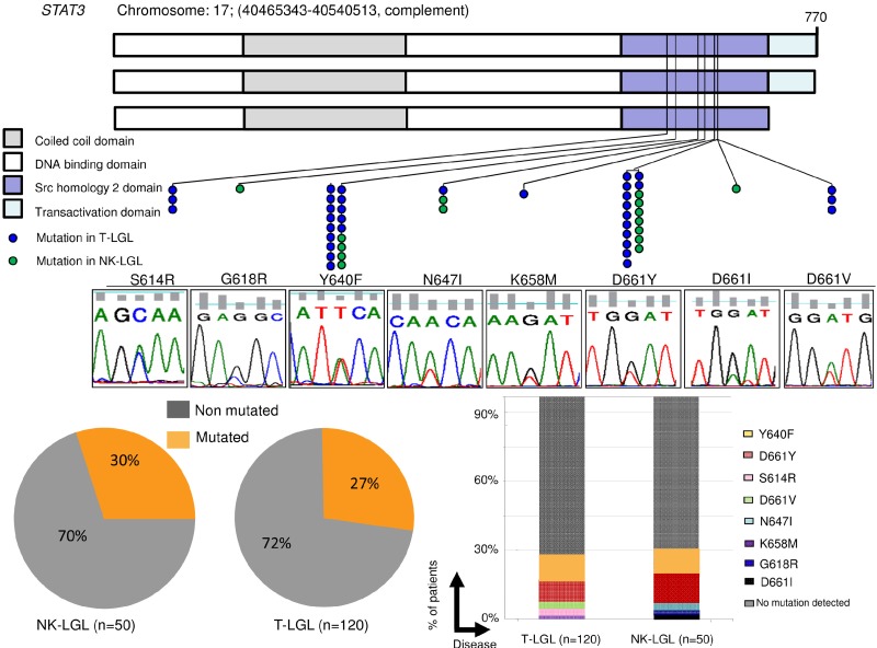
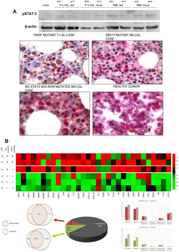
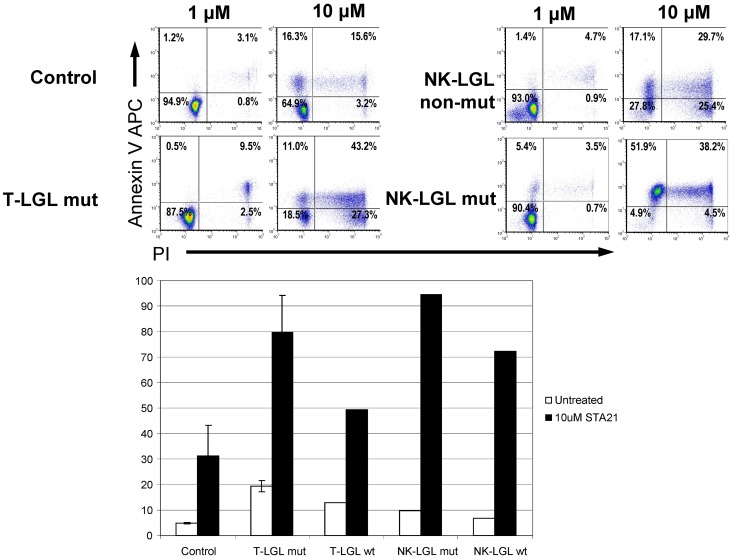
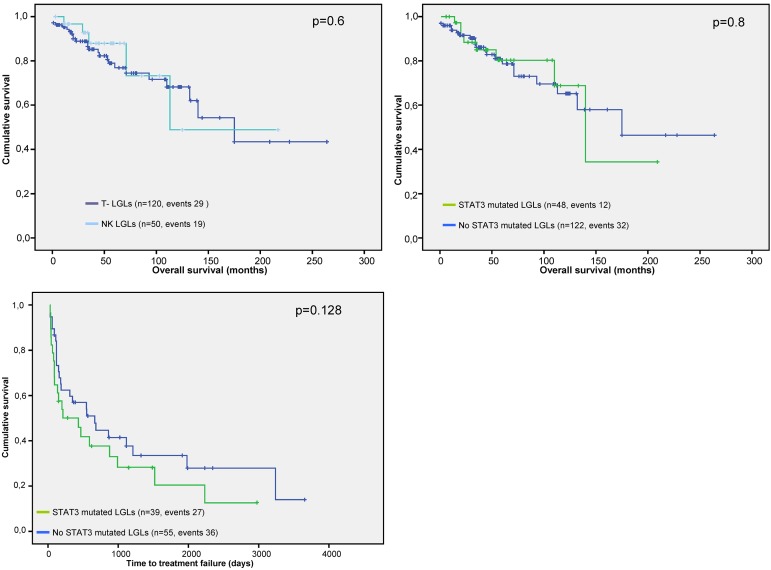
Chronic lymphoproliferative disorders of natural killer cells (CLPD-NKs) and T-cell large granular lymphocytic leukemias (T-LGLs) are clonal lymphoproliferations arising from either natural killer cells or cytotoxic T lymphocytes (CTLs). We have investigated for distribution and functional significance of mutations in 50 CLPD-NKs and 120 T-LGL patients by direct sequencing, allele-specific PCR, and microarray analysis. STAT3 gene mutations are present in both T and NK diseases: approximately one-third of patients with each type of disorder convey these mutations. Mutations were found in exons 21 and 20, encoding the Src homology 2 domain. Patients with mutations are characterized by symptomatic disease (75%), history of multiple treatments, and a specific pattern of STAT3 activation and gene deregulation, including increased expression of genes activated by STAT3. Many of these features are also found in patients with wild-type STAT3, indicating that other mechanisms of STAT3 activation can be operative in these chronic lymphoproliferative disorders. Treatment with STAT3 inhibitors, both in wild-type and mutant cases, resulted in accelerated apoptosis. STAT3 mutations are frequent in large granular lymphocytes suggesting a similar molecular dysregulation in malignant chronic expansions of NK and CTL origin. STAT3 mutations may distinguish truly malignant lymphoproliferations involving T and NK cells from reactive expansions.


Figures

Figure 1
Figure 1
Distribution of STAT3 mutations throughout gene domains and patient cohort. (A) STAT3 mutations (blue dots represent mutations in T-LGL; green dots, in CLPD-NKs) found in the SH2 domain, necessary for receptor association and tyrosine phosphodimer formation. The major domains of STAT3 are shown: coiled-coil domain, DNA-binding domain, SH2 domain, and transactivation domain. Lower panel: Corresponding representative Sanger sequence for each mutation found. (B) Percentage of patients with STAT3 mutations. Lesions were observed in 15 of 50 CLPD-NKs and 33 of 120 T-LGL patients when using Sanger and AS-PCR (7 cases not detected by Sanger). (C) Histograms showing the percentage of cases corresponding to each mutation. D661Y and Y640F accounted for ∼ 80% of all mutations found.
Figure 2
Figure 2
Proliferation and survival signals in chronic LGL diseases. (A) Constitutive STAT3 activation in leukemic cells. (Ai) Western blot analysis in leukemic cells from 4 T-LGL patients (2 mutated and 2 wild-type), 4 CLPD-NK patients (2 mutated and 2 wild-type), and a control. (Aii) Aberrant intracellular pSTAT3 signal (brown) has been also detected in paraffin sections from bone marrow biopsy samples of STAT3-mutant and nonmutant cases of T and NK origin. Previous immunohistochemical staining with CD8, surface CD3, and CD2 defined the cell lineage of the lymphocyte infiltration. Positive double staining with anticytoplasmic CD3 (pink) and pSTAT3 (brown) showed aberrant pSTAT3 signal in the infiltrating lymphoid compartment. Finally, a healthy donor tonsil sample shows no brown nuclei in cCD3-positive cells. (B) STAT3 pathway-related genes deregulated. (Bi) Heat map reflecting color-coded expression levels from a set of genes known to be regulated by STAT3 (columns) in purified T-LGL cells from 3 patients and control samples (rows). (Bii) Pie chart depicting whole genome expression in the 3 T-LGL leukemia patients and overlapping circles showing a high degree of coincidences in deregulated genes in mutated and nonmutated patients. (Ciii) Histograms of whole genome expression levels separated by pathways, exposing a predominance of deregulation in apoptosis and cell death, both in mutated and nonmutated patients. Up-regulated pathway genes are shown in pink (top panel) and down-regulated pathway genes in green (bottom panel).
Figure 3
Figure 3
Effect of STA-21 on apoptosis of malignant LGLs. Leukemic and control cells were harvested after 48 hours of STA-21 treatment and analyzed with propidium iodide and annexin V staining assays. (Top) Dose-dependent increase in apoptosis. (Bottom) Histograms depicting percentage of cells undergoing apoptosis after treatment compared with untreated cells.
Figure 4
Figure 4
Marked immunodominant Vβ expansions can be seen both in STAT3 SH2 domain-mutated (orange bars) and nonmutated patients (black bars).
Figure 5
Figure 5
Survival outcomes and time-to-treatment failure in patients with CLPD-NKs and T-LGL. P values presented correspond to the Cox regression between the groups indicated. (A) Comparison of survival outcomes according to the leukemic cell lineage. (B) Comparison of survival outcomes depending on the STAT3 SH2 domain mutational status. (C) Differences in the time-to-treatment failure in patients with or without STAT3 SH2 domain mutation. Time-to-treatment failure was defined as the interval between the start of treatment and the need for initiating a second line of therapy and/or progressive disease (including relapse after remission).

References

    1. Loughran TP, Jr, Kadin ME, Starkebaum G, et al. Leukemia of large granular lymphocytes: association with clonal chromosomal abnormalities and autoimmune neutropenia, thrombocytopenia, and hemolytic anemia. Ann Intern Med. 1985;102(2):169–175. - PubMed
    1. Loughran TP., Jr Clonal diseases of large granular lymphocytes. Blood. 1993;82(1):1–14. - PubMed
    1. Swerdlow SH. WHO Classification of Tumours of Haematopoietic and Lymphoid Tissues. 4th Ed. Lyon, France: International Agency for Research on Cancer; 2008. International Agency for Research on Cancer, World Health Organization.
    1. Swerdlow SH, Harris NL, editors. WHO Classification of Tumours of Haematopoietic and Lymphoid Tissues. 4th Ed. Lyon, France: International Agency for Research on Cancer; 2008.
    1. Bareau B, Rey J, Hamidou M, et al. Analysis of a French cohort of patients with large granular lymphocyte leukemia: a report on 229 cases. Haematologica. 2010;95(9):1534–1541. - PMC - PubMed

Publication types

MeSH terms