. 2012 Aug 17;337(6096):839-42.
doi: 10.1126/science.1222826. Epub 2012 Aug 2.

Circadian rhythm of redox state regulates excitability in suprachiasmatic nucleus neurons

Affiliations

Circadian rhythm of redox state regulates excitability in suprachiasmatic nucleus neurons

Tongfei A Wang et al. Science. .

Abstract

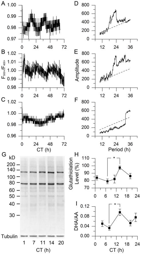
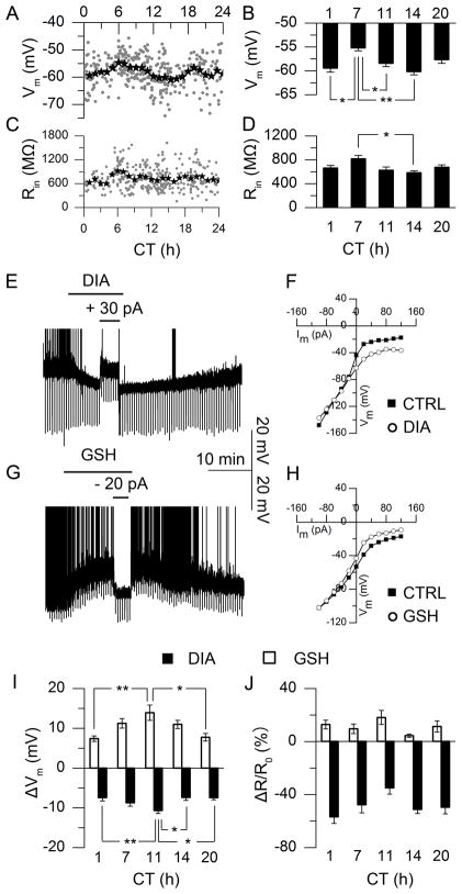
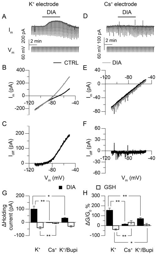
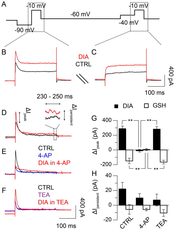
Daily rhythms of mammalian physiology, metabolism, and behavior parallel the day-night cycle. They are orchestrated by a central circadian clock in the brain, the suprachiasmatic nucleus (SCN). Transcription of clock genes is sensitive to metabolic changes in reduction and oxidation (redox); however, circadian cycles in protein oxidation have been reported in anucleate cells, where no transcription occurs. We investigated whether the SCN also expresses redox cycles and how such metabolic oscillations might affect neuronal physiology. We detected self-sustained circadian rhythms of SCN redox state that required the molecular clockwork. The redox oscillation could determine the excitability of SCN neurons through nontranscriptional modulation of multiple potassium (K(+)) channels. Thus, dynamic regulation of SCN excitability appears to be closely tied to metabolism that engages the clockwork machinery.


Figures

Fig. 1
Fig. 1
Circadian oscillation of redox state in rodent SCN. (A–C) Real-time imaging of relative redox state in SCN of wild-type (WT) rat (A), WT mouse (B) and Bmal1 −/− mouse (C). (D–F) χ2 periodograms (solid) of redox oscillations in SCN of WT rat (D), WT mouse (E), and Bmal1 −/− mouse (F), based on data in A–C, respectively, with the confidence interval of 0.001 (dashed); τrat = 23.74 ± 0.26 h (mean ± SD), τmouse = 23.75 ± 0.30 h; N = 5 for each group. (G) Glutathiolation patterns of BioGEE incorporation into rat SCN tissue over 5 points of circadian time (CT, which has a free-running time-base driven by the endogenous clock). (H) Protein glutathiolation over 5 CTs in rat SCN (P < 0.05, One-Way ANOVA; *, P < 0.05, Tukey’s Honestly Significant Difference (HSD) Test; N = 6). (I) DHA/AA ratio in rat SCN over 5 CTs (P < 0.05, One-Way ANOVA; *, P < 0.05, Tukey’s HSD Test; N = 3).
Fig. 2
Fig. 2
Circadian oscillations of neuronal excitability and redox regulation in rat SCN neurons. (A) Individual neurons (grey dots, 2-min recording, N = 364) and 1-h averages (star) of membrane potential (Vm). (B) Vm means at 5 CTs (P < 0.001, One-Way ANOVA; **, P < 0.01, *, P < 0.05, Tukey’s HSD Test; N = 36–60). (C) Membrane input resistance (Rin) measured by hyperpolarizing current steps (N = 337). (D) Average Rin from I–V constructed by current steps from −100 to +120 pA (20 pA increments, 800 ms duration) at 5 CTs (P < 0.05, One-Way ANOVA; *, P < 0.05, Tukey’s HSD Test; N = 15–30). (E, G) Current-clamp recording of Vm in response to oxidizing (E, diamide, DIA, 5 mM) or reducing reagent (G, glutathione, GSH, 1 mM), truncated spontaneous action potentials (SAP). (F, H) I–V curve before (filled) and during (open) DIA (F) or GSH (H) treatment. (I) Redox-induced ΔVm at 5 CTs (P < 0.01, One-Way ANOVA; **, P < 0.01, *, P < 0.05, Tukey’s HSD Test; N = 10–20). (J) Redox-induced ΔRin/Rin0% at 5 CTs (P > 0.05, One-Way ANOVA; N = 5–9).
Fig. 3
Fig. 3
Redox regulation of neuronal excitability through K+ current (CT 9–13). (A) Voltage-clamp recording of SCN neuron, with repeating slow-ramped voltage commands from −50 mV to −110 mV. (B) I–V curve constructed based on the command voltage and membrane current recorded, before (black) and during (grey) DIA treatment. (C) The DIA-evoked current as calculated from the difference in the membrane response in (B). (D–F) Similar voltage clamp recordings as (A–C), except that Cs+ replaced K+ in the patch pipette. (G, H) Holding current (G) and conductance (H) changes induced by redox reagents (DIA, black; GSH, white) with electrodes containing K+ (P < 0.01, paired Student’s t-Test to control; N = 6), Cs+ (P > 0.05 (current), P < 0.01 (conductance) for DIA, P < 0.05 for GSH, paired Student’s t-Test to control; N = 6), or K+ (in electrode) with bupivacaine (Bupi, 100 μM) in bath (P < 0.01, paired Student’s t-Test to control; N = 5); for the comparison across groups, P < 0.01, One-Way ANOVA; **, P < 0.01, *, P < 0.05, Tukey’s HSD Test.
Fig. 4
Fig. 4
Redox regulation of voltage-dependent K+ currents (CT 9 – 13). (A) Recording protocol of repeating voltage-step commands to voltage-clamped SCN neurons (15), before, during, and after drug treatment. (B, C) Current responses to the voltage-step commands of −10 mV pulses, following either −90 mV (B) or −40 mV (C) pre-pulse, before (black) or during (red) DIA treatment. (D) Voltage-dependent outward current in response to −10 mV voltage-step stimulation, calculated from the difference between the current responses in (B) and (C). (E, F) Effects of 4-aminopyridine (4-AP, 5 mM) and tetraethylammonium (TEA, 20 mM) on outward current evoked by DIA. (G) Transient current (< 10 ms) changes in response to redox treatment (DIA, black; GSH, white), with or without 4-AP or TEA (P < 0.01, One-Way ANOVA; **, P < 0.01, Tukey’s HSD Test; N = 5–6). (H) Persistent current (230–250 ms) changes in response to redox treatment, with/without 4-AP or TEA (P > 0.05, One-Way ANOVA; N = 5–6).

Comment in

  • Physiology. Circadian time redoxed.
    Belle MD, Piggins HD. Belle MD, et al. Science. 2012 Aug 17;337(6096):805-6. doi: 10.1126/science.1227203. Science. 2012. PMID: 22904000 No abstract available.

References

    1. Green CB, Takahashi JS, Bass J. The meter of metabolism. Cell. 2008;134:728. - PMC - PubMed
    1. Bass J, Takahashi JS. Circadian integration of metabolism and energetics. Science. 2010;330:1349. - PMC - PubMed
    1. Lowrey PL, Takahashi JS. Mammalian circadian biology: elucidating genome-wide levels of temporal organization. Annu Rev Genomics Hum Genet. 2004;5:407. - PMC - PubMed
    1. Prosser RA, Gillette MU. Cyclic changes in cAMP concentration and phosphodiesterase activity in a mammalian circadian clock studied in vitro. Brain Res. 1991;568:185. - PubMed
    1. O’Neill JS, Maywood ES, Chesham JE, Takahashi JS, Hastings MH. cAMP-dependent signaling as a core component of the mammalian circadian pacemaker. Science. 2008;320:949. - PMC - PubMed

Publication types