. 2012 Sep;23(9):1506-17.
doi: 10.1681/ASN.2012010079. Epub 2012 Aug 2.

Aquaporin 2 promotes cell migration and epithelial morphogenesis

Affiliations

Aquaporin 2 promotes cell migration and epithelial morphogenesis

Ying Chen et al. J Am Soc Nephrol. 2012 Sep.

Abstract

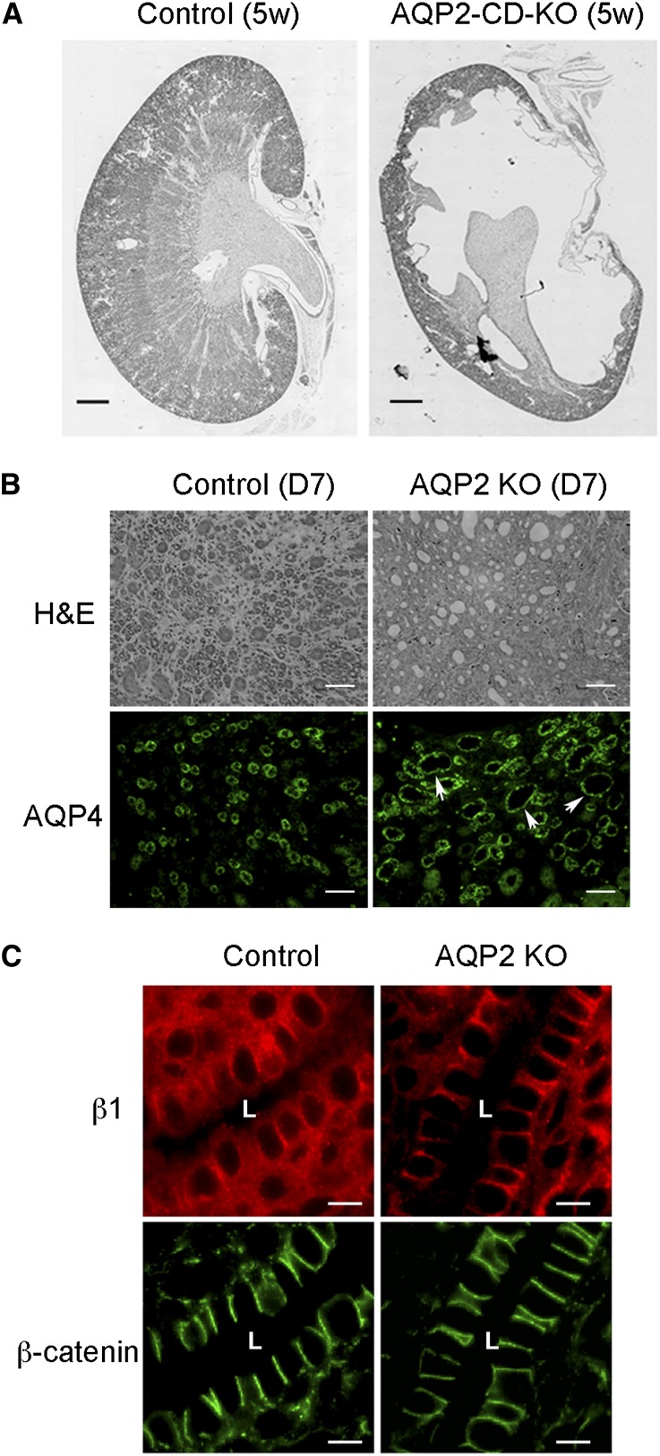
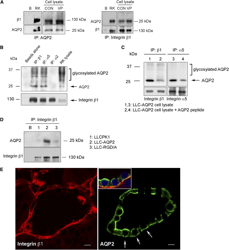
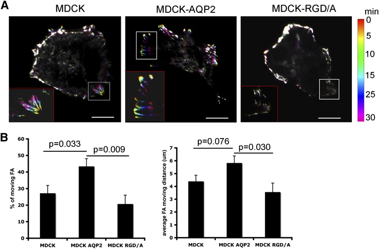
The aquaporin 2 (AQP2) water channel, expressed in kidney collecting ducts, contributes critically to water homeostasis in mammals. Animals lacking or having significantly reduced levels of AQP2, however, have not only urinary concentrating abnormalities but also renal tubular defects that lead to neonatal mortality from renal failure. Here, we show that AQP2 is not only a water channel but also an integrin-binding membrane protein that promotes cell migration and epithelial morphogenesis. AQP2 expression modulates the trafficking and internalization of integrin β1, facilitating its turnover at focal adhesions. In vitro, disturbing the interaction between AQP2 and integrin β1 by mutating the RGD motif led to reduced endocytosis, retention of integrin β1 at the cell surface, and defective cell migration and tubulogenesis. Similarly, in vivo, AQP2-null mice exhibited significant retention of integrin β1 at the basolateral membrane and had tubular abnormalities. In summary, these data suggest that the water channel AQP2 interacts with integrins to promote renal epithelial cell migration, contributing to the structural and functional integrity of the mammalian kidney.


Figures

Figure 1.
Figure 1.
Tubular abnormalities and altered distribution of integrin β1 in AQP2 knockout animals. (A) Gross abnormalities in kidney structure in AQP2 knockout animals 5 weeks after birth. (B) Tubular dilation and microcyst formation (arrows) in AQP2 knockout animals at postnatal day 7. AQP4 (green) indicates the tubules are collecting ducts. (C) Altered distribution of integrin β1 in collecting ducts of AQP2 knockout animals. Integrin β1 (red) is in the sub-basal membrane domain in wild-type (control) animals, but is concentrated on the lateral membrane in knockout mice. The subcellular distribution of β-catenin (green) is not altered. KO, knockout; L, lumen. Scale bar, 1 mm in A; 50 μm in B; 10 μm in C.
Figure 2.
Figure 2.
AQP2 interacts with integrins via an RGD motif. (A) AQP2 and integrin β1 coimmunoprecipitate (co-IP) from rat kidney (RK) and cell lysates (from stable LLCPK1 cells expressing wild-type AQP2), using anti-AQP2 (left) and anti-integrin β1 (right) antibodies for IP. Co-IP was also performed in vasopressin-stimulated cells (vasopressin). The presence of AQP2 or integrin β1 in corresponding co-IP complex is shown in the lower panel. (B) AQP2 interacts with integrin α5, but not integrin α2 by co-IP using RK lysate. Integrin β1 is present in the co-IP complex using integrin α2, α5, and β1 antibodies for IP and is shown in lower panel. (C) Synthetic AQP2 peptide attenuates the interaction of AQP2 and integrin β1, and AQP2 and integrin α5. (D) Mutation of the AQP2 RGD motif (lane 3, AQP2 RGD/A-expressing LLCPK1 cells) attenuates AQP2 and integrin β1 interaction. Data are representative of at least three experiments. “B” indicates beads only. (E) Immunofluorescence staining reveals the co-presence of AQP2 (green, arrow) and integrin β1 signal (red) in the basal membrane domain in tubular cells of the collecting ducts in RK. The insert in right panel highlights the colocalization of AQP2 and integrin β1 on the basal membrane of the AQP2-expressing principal cells. Scale bar, 10 μm.
Figure 3.
Figure 3.
AQP2 promotes kidney epithelial cell migration. (A) The expression of endogenous canine AQP2 in untransfected MDCK cells is detected by RT-PCR using dog specific AQP2 primer (dAQP2) and the expression of transfected rat AQP2 is detected by RT-PCR using rat specific AQP2 primer (rAQP2). The presence of endogenous AQP2 in MDCK cells is also detected by immunoblot after overloading the protein lysates of MDCK cells. The expression of integrins β1, α2, and α5 is equal among these cells. (B) AQP2 (green) is polarized to the lamellipodia/leading edge and colocalizes with integrin β1 (red) in the lamellipodia (arrow head) in migrating MDCK cells expressing wild-type AQP2 (upper panel). However, AQP2 does not polarize to the lamellipodia and neither does it colocalize with integrin β1 in the lamellipodia in MDCK cells expressing AQP2 RGD/A mutant (lower panel). Arrow indicates the direction of cell migration. (C) Expression of AQP2 (middle panel) promotes, whereas expression of AQP2 RGD/A inhibits cell migration in a wound-healing assay on fibronectin-coated surface compared with untransfected MDCK cells. The average speed of cell migration over 15 hours was calculated and shown in the right penal. Values are mean ± SEM, n=3, *P<0.05, **P<0.01. Scale bar, 5 μm in B.
Figure 4.
Figure 4.
AQP2 modulates the trafficking and subcellular distribution of integrin β1. (A) Expressing wild-type AQP2 in MDCK cells facilitates, whereas a lack of AQP2 expression or expression of AQP2 RGD/A inhibits the internalization of surface-labeled integrin β1 (red) over time. (B) Ratios of membrane retained integrin β1 signal over total integrin β1 signal (membrane and cytosol) after integrin β1 internalization over time were plotted. Values represent the mean ± SEM from at least 50 cells at each time point from three independent experiments. Membrane retention of surface-labeled integrin β1 was significant at 60 and 120 minutes after internalization in untransfected MDCK cells compared with cells expressing wild-type AQP2. A more dramatic membrane accumulation is seen in MDCK cells overexpressing AQP2 RGD/A. (C) Overlap of surface-labeled, internalized β1 integrin signal (blue) with AQP2 (red) and GFP-clathrin (green, upper panel) as well as GFP-rab11 (green, lower panel) is observed in vesicles. Scale bar, 5 μm in A; 2 μm in C.
Figure 5.
Figure 5.
AQP2 expression modulates the cell surface expression of integrin β1. (A) AQP2 expression (middle) attenuates membrane accumulation of integrin β1 (red) in polarized MDCK cells compared with untransfected cells (left), whereas expression of AQP2 RGD/A (right) causes extensive lateral membrane accumulation of integrin β1. (B) Cell surface retention of integrin β1 and integrin α5 in untransfected cells (MDCK) and more dramatically in AQP2 RGD/A (D/A)-expressing cells was confirmed by surface biotinylation. Near equal input of total cellular protein is shown by immunoblot using glyceraldehyde 3-phosphate dehydrogenase. No difference of the surface expression of integrin α2 or transferrin receptor was found. The amount of biotinylated integrin β1, α5, and α2 in AQP2 and AQP2 RGD/A-expressing cells was normalized to untransfected MDCK cells (control). Data are presented as mean ± SEM, n=4. n.s, no significant difference.
Figure 6.
Figure 6.
AQP2 modulates the dynamics of FAs on fibronectin-coated surface. (A) Expression of AQP2 in MDCK cells promotes mobilization of GFP-integrin β1 in FAs by TIRF microscopy. Images taken at 0, 5, 10, 20, and 30 minutes are encoded red, yellow, green, blue, and purple, respectively in rainbow-color overlaid images. A representative moving region of FAs from individual cells is highlighted and enlarged in the left corner of each image. (B) The percentage of motile FAs and the average moving distance of individual FA was quantified. n=412–440 FAs from 7 to 9 cells for each cell line. Values are presented as mean ± SEM. Data were obtained from at least three independent experiments. Scale bar, 5 μm in A.

Comment in

  • Aquaporin 2: not just for moving water.
    Sands JM. Sands JM. J Am Soc Nephrol. 2012 Sep;23(9):1443-4. doi: 10.1681/ASN.2012060613. Epub 2012 Jul 12. J Am Soc Nephrol. 2012. PMID: 22797179 Free PMC article. No abstract available.

References

    1. Brown D: The ins and outs of aquaporin-2 trafficking. Am J Physiol Renal Physiol 284: F893–F901, 2003 - PubMed
    1. Lorenz D, Krylov A, Hahm D, Hagen V, Rosenthal W, Pohl P, Maric K: Cyclic AMP is sufficient for triggering the exocytic recruitment of aquaporin-2 in renal epithelial cells. EMBO Rep 4: 88–93, 2003 - PMC - PubMed
    1. van Balkom BW, Graat MP, van Raak M, Hofman E, van der Sluijs P, Deen PM: Role of cytoplasmic termini in sorting and shuttling of the aquaporin-2 water channel. Am J Physiol Cell Physiol 286: C372–C379, 2004 - PubMed
    1. Sun TX, Van Hoek A, Huang Y, Bouley R, McLaughlin M, Brown D: Aquaporin-2 localization in clathrin-coated pits: Inhibition of endocytosis by dominant-negative dynamin. Am J Physiol Renal Physiol 282: F998–F1011, 2002 - PubMed
    1. Lu H, Sun TX, Bouley R, Blackburn K, McLaughlin M, Brown D: Inhibition of endocytosis causes phosphorylation (S256)-independent plasma membrane accumulation of AQP2. Am J Physiol Renal Physiol 286: F233–F243, 2004 - PubMed

Publication types

MeSH terms