

Alterations in periarticular bone and cross talk between subchondral bone and articular cartilage in osteoarthritis
- PMID: 22859924
- PMCID: PMC3403248
- DOI: 10.1177/1759720X12437353
Alterations in periarticular bone and cross talk between subchondral bone and articular cartilage in osteoarthritis
Abstract
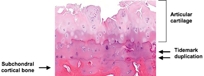
The articular cartilage and the subchondral bone form a biocomposite that is uniquely adapted to the transfer of loads across the diarthrodial joint. During the evolution of the osteoarthritic process biomechanical and biological processes result in alterations in the composition, structure and functional properties of these tissues. Given the intimate contact between the cartilage and bone, alterations of either tissue will modulate the properties and function of the other joint component. The changes in periarticular bone tend to occur very early in the development of OA. Although chondrocytes also have the capacity to modulate their functional state in response to loading, the capacity of these cells to repair and modify their surrounding extracellular matrix is relatively limited in comparison to the adjacent subchondral bone. This differential adaptive capacity likely underlies the more rapid appearance of detectable skeletal changes in OA in comparison to the articular cartilage. The OA changes in periarticular bone include increases in subchondral cortical bone thickness, gradual decreases in subchondral trabeular bone mass, formation of marginal joint osteophytes, development of bone cysts and advancement of the zone of calcified cartilage between the articular cartilage and subchondral bone. The expansion of the zone of calcified cartilage contributes to overall thinning of the articular cartilage. The mechanisms involved in this process include the release of soluble mediators from chondrocytes in the deep zones of the articular cartilage and/or the influences of microcracks that have initiated focal remodeling in the calcified cartilage and subchondral bone in an attempt to repair the microdamage. There is the need for further studies to define the pathophysiological mechanisms involved in the interaction between subchondral bone and articular cartilage and for applying this information to the development of therapeutic interventions to improve the outcomes in patients with OA.
Keywords: articular cartilage; biomechanics; bone remodeling; osteoarthritis.
Conflict of interest statement
Figures
References
-
- Amin A.K., Huntley J.S., Simpson A.H., Hall A.C. (2009) Chondrocyte survival in articular cartilage: the influence of subchondral bone in a bovine model. J Bone Joint Surg Br 91(5): 691–699 - PubMed
-
- Ashraf S., Mapp P.I., Walsh D.A. (2011) Contributions of angiogenesis to inflammation, joint damage and pain in a rat model of osteoarthritis. Arthritis Rheum, in press - PubMed
-
- Bancroft L.W., Peterson J.J., Kransdorf M.J. (2004) Cysts, geodes, and erosions. Radiol Clin North Am 42: 73–87 - PubMed
-
- Bau B., Gebhard P.M., Haag J., Knorr T., Bartnik E., Aigner T. (2002) Relative messenger RNA expression profiling of collagenases and aggrecanases in human articular chondrocytes in vivo and in vitro. Arthritis Rheum 46: 2648–2657 - PubMed
-
- Bennell K.L., Creaby M.W., Wrigley T.V., Bowles K.A., Hinman R.S., Cicuttini F., et al. (2011) Bone marrow lesions are related to dynamic knee loading in medial knee osteoarthritis. Ann Rheum Dis 69: 1151–1154 - PubMed
LinkOut - more resources
Full Text Sources
Other Literature Sources
Medical
