. 2012 Aug;4(4):301-11.
doi: 10.1177/1759720X11436240.

Imaging in ankylosing spondylitis

Affiliations

Imaging in ankylosing spondylitis

Mikkel Ostergaard et al. Ther Adv Musculoskelet Dis. 2012 Aug.

Abstract

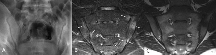
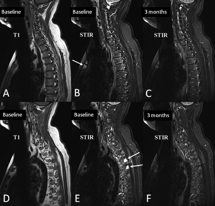
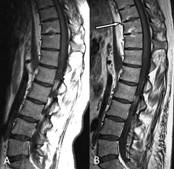
Imaging is an integral part of the management of patients with ankylosing spondylitis and axial spondyloarthritis. Characteristic radiographic and/or magnetic resonance imaging (MRI) findings are key in the diagnosis. Radiography and MRI are also useful in monitoring the disease. Radiography is the conventional, albeit quite insensitive, gold standard method for assessment of structural damage in spine and sacroiliac joints, whereas MRI has gained a decisive role in monitoring disease activity in clinical trials and practice. MRI may also, if ongoing research demonstrates a sufficient reliability and sensitivity to change, become a new standard method for assessment of structural damage. Ultrasonography allows visualization of peripheral arthritis and enthesitis, but has no role in the assessment of axial manifestations. Computed tomography is a sensitive method for assessment of structural changes in the spine and sacroiliac joints, but its clinical utility is limited due to its use of ionizing radiation and lack of ability to assess the soft tissues. It is exciting that with continued dedicated research and the rapid technical development it is likely that even larger improvements in the use of imaging may occur in the decade to come, for the benefit of our patients.

Keywords: ankylosing spondylitis; computed tomography; imaging; magnetic resonance imaging; radiography; spondyloarthritis; ultrasonography; ultrasound.


Conflict of interest statement

Conflict of interest statement: The authors declare no conflicts of interest in preparing this article.

Figures

Figure 1.
Figure 1.
Radiographic findings in sacroiliac joints and spine in ankylosing spondylitis. (A) Radiograph of the sacroiliac joints in a 23-year-old male demonstrates established ankylosing spondylitis. Bilateral erosions cause discrete foci of loss of subchondral bone and apparent joint space widening in some areas (arrows) and ill definition of the joint margin in other areas (arrowheads). Bilateral subchondral sclerosis is most prominent in the left ilium. (B)–(D) Radiographs of the spine in a 47-year-old male with widespread ankylosis. The cervical spine (B) exhibits extensive formation of vertical syndesmophytes that have bridged the anterior vertebral corners causing ankylosis. Some facet joints are fused, best appreciated at C2/3. The lumbar spine (C; enlargement of L1–L4 in D) shows similar ankylosis. Note the thick right L1/2 bridging (arrow) and compare this to the delicate vertical syndesmophytosis on the left at L3/4. The sacroiliac joints are completely fused with barely any remnant of joint visible.
Figure 2.
Figure 2.
Early sacroiliitis on conventional radiography and MRI. Radiograph (A) of the sacroiliac joints in a 28-year-old male reveals only subtle findings of possible erosion and minimal sclerosis. Short tau inversion recovery (STIR) MRI image (B) performed at the same time shows multiple bone marrow lesions, which appear as oedema (bright; arrows) involving the sacrum and ilium bilaterally, i.e. definite sacroiliitis was documented by MRI. The corresponding T1-weighted MRI image (C) shows some areas of diminished marrow fat signal corresponding to the intense oedema in the left upper quadrant. Some very subtle defects in the subchondral marrow in the lower quadrants, which likely represent tiny erosions (arrows), are also seen.
Figure 3.
Figure 3.
Inflammatory and fat lesions on MRI of the spine. MRI of the lumbar and lower thoracic spine in 27-year-old male shows multiple tiny foci of infiltration of fat in the posterior corners of vertebral bodies on the T1-weighted sequence (A; arrows). On the short tau inversion recovery (STIR) sequence (B), these discs demonstrate no evidence of degeneration of the nucleus pulposis or tear of the annulus fibrosus, which is consistent with a postinflammatory cause of the marrow fat deposition rather than trauma or degenerative disc disease. Also note the solitary focus of inflammation on STIR imaging with increased signal at the anterosuperior corner of T10 (arrowhead). The appearance is typical for a corner inflammatory lesion (CIL) associated with spondyloarthritis (i.e. a triangular shaped lesion which may or may not (as in this case) be quite as bright in the extreme corner, with adjacent normal nucleus pulposus).
Figure 4.
Figure 4.
Bone marrow oedema in the transverse processes, costovertebral joints and manubriosternal joint. MRI of the cervical and upper thoracic spine of a 29-year-old male patient scans were performed before (A–B and D–E) and 3 months after (C and F) initiation of anti-TNF therapy. Sagittal slices lateral to the spinal canal are shown. (A)–(C) Sagittal slice through the pedicle and the lateral parts of the vertebrae, shows moderate bone oedema on the baseline STIR image (B) in the posterolateral aspects of all of the thoracic vertebral bodies, most pronounced at T4 and T5. The distribution is typical for inflammation on the vertebral side of the costovertebral joints. Also note inflammation in the manubriosternal joint anteriorly (arrow). Most foci of inflammation are still faintly visible after anti-TNF therapy (C), but are clearly less intense. (D)–(F) Far lateral slice through the transverse processes and ribs. Intense bone marrow oedema is seen in the transverse processes of the upper thoracic spine (E; arrows), which resolves completely with treatment. This pattern of bone marrow oedema in the transverse processes, costovertebral joints and manubriosternal joint are pathognomonic of spondyloarthritis.
Figure 5.
Figure 5.
Progression of structural damage in the spine in ankylosing spondylitis, visualized by MRI. T1-weighted MRIs of the thoracolumbar spine with a 3-year interval in a baseline (A) 23-year-old male with established ankylosing spondylitis. The follow-up MRI (B) demonstrates new anterior ankylosis at T9/10 with bridging anterior syndesmophytosis containing marrow fat signal. Note also several other findings indicating the progression of structural damage: the intervertebral disc at T10/11 is completely fused on the second scan; a new endplate defect has appeared at the inferior endplate of T9 with new fat infiltration; and the signal within the T8/9 disc has increased suggesting progression of disc ossification.

Similar articles

Cited by

References

    1. Baraliakos X., Hermann K.G., Landewe R., Listing J., Golder W., Brandt J., et al. (2005) Assessment of acute spinal inflammation in patients with ankylosing spondylitis by magnetic resonance imaging: a comparison between contrast enhanced T1 and short tau inversion recovery (STIR) sequences. Ann Rheum Dis 64: 1141–1144 - PMC - PubMed
    1. Baraliakos X., Listing J., Rudwaleit M., Sieper J., Braun J. (2008) The relationship between inflammation and new bone formation in patients with ankylosing spondylitis. Arthritis Res Ther 10: R104. - PMC - PubMed
    1. Bennett A.N., McGonagle D., O’Connor P., Hensor E.M., Sivera F., Coates L.C., et al. (2008) Severity of baseline magnetic resonance imaging-evident sacroiliitis and HLA-B27 status in early inflammatory back pain predict radiographically evident ankylosing spondylitis at eight years. Arthritis Rheum 58: 3413–3418 - PubMed
    1. Berens D.L. (1971) Roentgen features of ankylosing spondylitis. Clin Orthop Relat Res 74: 20–33 - PubMed
    1. Braun J., Baraliakos X., Golder W., Brandt J., Rudwaleit M., Listing J., et al. (2003) Magnetic resonance imaging examinations of the spine in patients with ankylosing spondylitis, before and after successful therapy with infliximab: evaluation of a new scoring system. Arthritis Rheum 48: 1126–1136 - PubMed