

Altered A-to-I RNA editing in human embryogenesis
- PMID: 22859999
- PMCID: PMC3409221
- DOI: 10.1371/journal.pone.0041576
Altered A-to-I RNA editing in human embryogenesis
Abstract
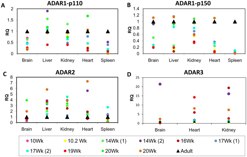
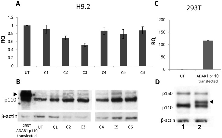
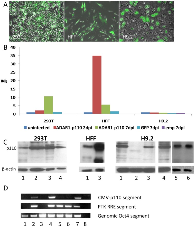
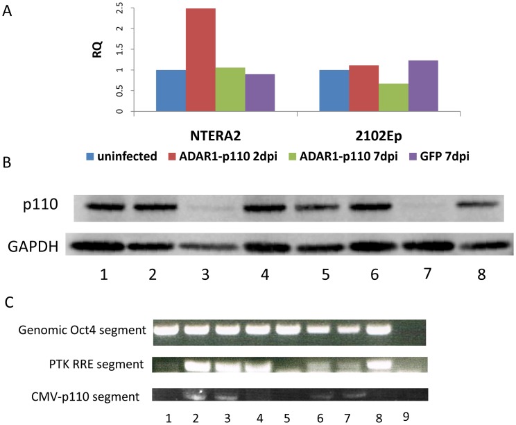
Post-transcriptional events play an important role in human development. The question arises as to whether Adenosine to Inosine RNA editing, catalyzed by the ADAR (Adenosine Deaminase acting on RNA) enzymes, differs in human embryogenesis and in adulthood. We tested the editing of various target genes in coding (FLNA, BLCAP, CYFIP2) and non-coding sequences at their Alu elements (BRCA1, CARD11, RBBP9, MDM4, FNACC), as well as the transcriptional levels of the ADAR1 enzymes. This analysis was performed on five fetal and adult human tissues: brain, heart, liver, kidney, and spleen, as well as on human embryonic stem cells (hESCs), which represent the blastocyst stage in early human development. Our results show substantially greater editing activity for most adult tissue samples relative to fetal ones, in six of the eight genes tested. To test the effect of reduced A-to-I RNA editing activity in early human development we used human embryonic stem cells (hESCs) as a model and tried to generate hESC clones that overexpress the ADAR1-p110 isoform. We were unable to achieve overexpression of ADAR1-p110 by either transfection or lentiviral infection, though we easily generated hESC clones that expressed the GFP transgene and overexpressed ADAR1-p110 in 293T cells and in primary human foreskin fibroblast (HFF) cells. Moreover, in contrast to the expected overexpression of ADAR1-p110 protein following its introduction into hESCs, the expression levels of this protein decreased dramatically 24-48 hr post infection. Similar results were obtained when we tried to overexpress ADAR1-p110 in pluripotent embryonal carcinoma cells. This suggests that ADAR1 protein is substantially regulated in undifferentiated pluripotent hESCs. Overall, our data suggest that A-to-I RNA editing plays a critical role during early human development.
Conflict of interest statement
Figures


Similar articles
-
Alu sequences in undifferentiated human embryonic stem cells display high levels of A-to-I RNA editing.PLoS One. 2010 Jun 21;5(6):e11173. doi: 10.1371/journal.pone.0011173. PLoS One. 2010. PMID: 20574523 Free PMC article.
-
Specificity of ADAR-mediated RNA editing in newly identified targets.RNA. 2008 Jun;14(6):1110-8. doi: 10.1261/rna.923308. Epub 2008 Apr 22. RNA. 2008. PMID: 18430892 Free PMC article.
-
Evolutionarily conserved human targets of adenosine to inosine RNA editing.Nucleic Acids Res. 2005 Feb 24;33(4):1162-8. doi: 10.1093/nar/gki239. Print 2005. Nucleic Acids Res. 2005. PMID: 15731336 Free PMC article.
-
A-to-I RNA editing and hematopoiesis.Exp Hematol. 2024 Nov;139:104621. doi: 10.1016/j.exphem.2024.104621. Epub 2024 Aug 24. Exp Hematol. 2024. PMID: 39187172 Review.
-
A-to-I editing of coding and non-coding RNAs by ADARs.Nat Rev Mol Cell Biol. 2016 Feb;17(2):83-96. doi: 10.1038/nrm.2015.4. Epub 2015 Dec 9. Nat Rev Mol Cell Biol. 2016. PMID: 26648264 Free PMC article. Review.
Cited by
-
Using mouse models to unlock the secrets of non-synonymous RNA editing.Methods. 2019 Mar 1;156:40-45. doi: 10.1016/j.ymeth.2018.10.016. Epub 2018 Oct 26. Methods. 2019. PMID: 30827465 Free PMC article. Review.
-
Adenosine Deaminase That Acts on RNA 3 (ADAR3) Binding to Glutamate Receptor Subunit B Pre-mRNA Inhibits RNA Editing in Glioblastoma.J Biol Chem. 2017 Mar 10;292(10):4326-4335. doi: 10.1074/jbc.M117.779868. Epub 2017 Feb 6. J Biol Chem. 2017. PMID: 28167531 Free PMC article.
-
Profiling A-to-I RNA editing during mouse somatic reprogramming at the single-cell level.Heliyon. 2023 Jul 13;9(7):e18133. doi: 10.1016/j.heliyon.2023.e18133. eCollection 2023 Jul. Heliyon. 2023. PMID: 37519753 Free PMC article.
-
An Evolutionary Landscape of A-to-I RNA Editome across Metazoan Species.Genome Biol Evol. 2018 Feb 1;10(2):521-537. doi: 10.1093/gbe/evx277. Genome Biol Evol. 2018. PMID: 29294013 Free PMC article.
-
Curcumin modulates the apolipoprotein B mRNA editing by coordinating the expression of cytidine deamination to uridine editosome components in primary mouse hepatocytes.Korean J Physiol Pharmacol. 2019 May;23(3):181-189. doi: 10.4196/kjpp.2019.23.3.181. Epub 2019 Apr 24. Korean J Physiol Pharmacol. 2019. PMID: 31080349 Free PMC article.
References
-
- Bass BL (1997) RNA editing and hypermutation by adenosine deamination. Trends Biochem Sci 22: 157–162. - PubMed
-
- Melcher T, Maas S, Herb A, Sprengel R, Higuchi M, et al. (1996) RED2, a brain-specific member of the RNA-specific adenosine deaminase family. J Biol Chem 271: 31795–31798. - PubMed
-
- Higuchi M, Maas S, Single FN, Hartner J, Rozov A, et al. (2000) Point mutation in an AMPA receptor gene rescues lethality in mice deficient in the RNA-editing enzyme ADAR2. Nature 406: 78–81. - PubMed
Publication types
MeSH terms
Substances
LinkOut - more resources
Full Text Sources
Research Materials
Miscellaneous
