. 2012;7(7):e42199.
doi: 10.1371/journal.pone.0042199. Epub 2012 Jul 30.

Subcellular localization of iron and heme metabolism related proteins at early stages of erythrophagocytosis

Affiliations

Subcellular localization of iron and heme metabolism related proteins at early stages of erythrophagocytosis

Constance Delaby et al. PLoS One. 2012.

Abstract

Background: Senescent red blood cells (RBC) are recognized, phagocytosed and cleared by tissue macrophages. During this erythrophagocytosis (EP), RBC are engulfed and processed in special compartments called erythrophagosomes. We previously described that following EP, heme is rapidly degraded through the catabolic activity of heme oxygenase (HO). Extracted heme iron is then either exported or stored by macrophages. However, the cellular localization of the early steps of heme processing and iron extraction during EP remains to be clearly defined.

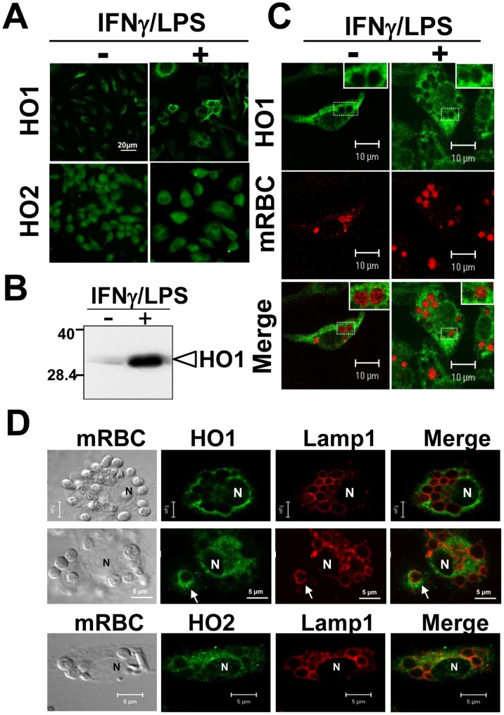
Methodology/principal findings: We took advantage of our previously described cellular model of EP, using bone marrow-derived macrophages (BMDM). The subcellular localization of both inducible and constitutive isoforms of HO (HO-1 and HO-2), of the divalent metal transporters (Nramp1, Nramp2/DMT1, Fpn), and of the recently identified heme transporter HRG-1, was followed by fluorescence and electron microscopy during the earliest steps of EP. We also looked at some ER [calnexin, glucose-6-phosphatase (G6Pase) activity] and lysosomes (Lamp1) markers during EP. In both quiescent and LPS-activated BMDM, Nramp1 and Lamp1 were shown to be strong markers of the erythrophagolysosomal membrane. HRG-1 was also recruited to the erythrophagosome. Furthermore, we observed calnexin labeling and G6Pase activity at the erythrophagosomal membrane, indicating the contribution of ER in this phagocytosis model. In contrast, Nramp2/DMT1, Fpn, HO-1 and HO-2 were not detected at the membrane of erythrophagosomes.

Conclusions/significance: Our study highlights the subcellular localization of various heme- and iron-related proteins during early steps of EP, thereby suggesting a model for heme catabolism occurring outside the phagosome, with heme likely being transported into the cytosol through HRG1. The precise function of Nramp1 at the phagosomal membrane in this model remains to be determined.


Conflict of interest statement

Competing Interests: The authors have declared that no competing interests exist.

Figures

Figure 1
Figure 1. Subcellular localization of Nramp1 protein during EP.
(A) IF staining of Nramp1 and Nramp2/DMT1 in untreated (−) or IFNγLPS-treated BMDM. Both Nramp1 and Nramp2/DMT1 expression are up-regulated after LPS/IFNγ treatment (B) IF of Nramp1 (left panels) was performed on quiescent Nramp1(+) or Nramp1(−) BMDM incubated with artificially-aged mRBC (1 hr). Nramp1 (+) or Nramp1 (−) BMDM present similar phagocytic activity as shown with hemoglobin autofluorescence at 520 nm (right panels) of the ingested mRBC. (C) Confocal microscopy analysis of the cellular distribution of Nramp1 (green) after EP in both quiescent (−) or LPS/IFNγ-treated BMDM. Ingested mRBC were visualized through auto-fluorescence of hemoglobin (red) or phase contrast. High magnifications (right panels) clearly show Nramp1 staining (green) at the phagosomal membrane surrounding ingested mRBC in both quiescent and activated BMDM (arrows). N: Nucleus.
Figure 2
Figure 2. Nramp1 but not Nramp2/DMT1 is recruited to the phagosomal membrane surrounding ingested mRBC.
After activation with LPS/IFNγ for 16 hrs, BMDM were processed for erythrophagocytosis assay (1 hr) and double IF labeling was performed as follows. (A and B): Nramp1 and Lamp1; (C): Nramp1 and TfR, (D): Nramp2/DMT1 and Lamp1; (E) Nramp2/DMT1 and TfR. Confocal analysis indicates that Nramp1 is present in vesicular endomembranes and is concentrated at the erythrophagosomal membrane where it colocalizes with Lamp1 (A and B) but displays a different localization than TfR (C). On the other hand Nramp2/DMT1 does not show any evidence of phagosomal membrane localization (D) but colocalizes with TfR (E) in recycling endosomes.
Figure 3
Figure 3. Heme oxygenase expression and localization in BMDM after activation and EP.
Expression of HO-1 and HO-2 was analyzed in quiescent (−) or activated (with LPS/IFNγ) BMDM by classical fluorescence (A) or by Western blot (B; for HO-1 only). HO-1 but not HO-2 was induced after pro-inflammatory cytokines treatment. (C) Localization of HO-1 during EP (1 hr) in quiescent or activated BMDM. Mouse RBC are visualized through autofluorescence of hemoglobin (middle panels). (D) Confocal analysis of HO proteins and Lamp1 staining during EP. HO-1 and HO-2 do not colocalize with Lamp1 at the phagosomal membrane but rather display a diffuse staining around the RBC-containing phagosome.
Figure 4
Figure 4. Endoplasmic reticulum and erythrophagocytosis.
BMDM were incubated with mouse artificially aged erythrocytes (mRBC) for 15 (A) to 30 min (B–G) and were processed either for calnexin fluorescence labeling (A) or G6Pase activity for EM analysis (B–G). In A, engulfed erythrocytes were visualized by phase contrast (upper panel) or by hemoglobin autofluorescence at 520 nm (middle panel). Lower panel in A shows calnexin labeling around one phagosome containing a RBC (arrow and inset). The nuclear membrane (N) and the ER (intracellular staining) are specifically labeled for calnexin. (B) At the ultrastrutural level, some ingested mRBC phagosomes were negative for G6Pase. A dense area located around the membrane of the phagosome was clearly negative for any cellular structure and for G6Pase activity (see inset high magnification). This region likely corresponds to a network of actin filaments surrounding the phagosome and indicates the early nature of this phagosome. In C, some G6Pase positive patches are organized around and close to the membrane of the mRBC-containing phagosome (see inset). In most of mRBC-containing phagosomes, EM sections reveal intense G6Pase labeling that accumulates between the phagosomal membrane and the membrane of the ingested erythrocyte (D to F; see arrows in inset). Some erythrophagosomes present irregular shapes associated with appearance of intra-phagosomal electron-opaque particles that likely correspond to G6Pase precipitates that diffuse throughout the phagosome (F and G; see results and discussion). In G (see inset), asterisks indicate vesicular compartments harboring strong G6Pase activity that may illustrate fragmentation of the erythrophagosome. The nuclear envelope (NE) and endoplasmic reticulum (ER) were specifically and strongly labeled.
Figure 5
Figure 5. HRG1 is recruited with Nramp1 to the phagosomal membrane surrounding ingested mRBC.
BMDM were processed for erythrophagocytosis assay (1 hr) and double IF labeling of HRG1 (red) and Nramp1 (green) was performed. Arrows indicate the position of erythrophagosomes. White bars in upper panels correspond to a size of 20 µm. In panel A, nuclei (N) are visualized with DAPI. Classical fluorescence (A) or confocal analysis (B) indicate that HRG1 and Nramp1 share similar localization within the cells. In addition, HRG1 and Nramp1 are both concentrated at the erythrophagosomal membrane (arrows).
Figure 6
Figure 6. Proposed model of heme iron recycling following EP of artificially-aged RBC.
Senescent RBC are specifically recognized and engulfed by BMDM inside an erythrophagosomal compartment. ER and lysosomes (LY) fuse with this compartment to form a phagolysosome (PL). During this process, Nramp1 is recruited at the phagosomal membrane. The exact role of Nramp1, strongly present at that site, still remains to be elucidated. Nramp2, primarily located in recycling endosomes, does not seem to be involved in this process. After hemoglobin degradation, heme is transported through the phagosomal membrane via HRG-1. Once in the cytosol, heme can exert its biological function and is then degraded by HO-1. Further investigations are necessary to clarify the subcellular site of heme catabolism by HO-1. Additionally, heme is transported outside the cell via FLVCR. Freed heme iron is then recycled to the circulation via Fpn or is stored in the form of cytosolic ferritin.

References

    1. Bratosin D, Mazurier J, Tissier JP, Estaquier J, Huart JJ, et al. (1998) Cellular and molecular mechanisms of senescent erythrocyte phagocytosis by macrophages. A review. Biochimie 80: 173–195. - PubMed
    1. Deiss A (1999) editor (1999) Destruction of erythrocytes. Lipincott Williams & Wilkins, Baltimore, MD. ed: Lee GR, Foerster J, Lukens J, Paraskevas F,Greer JP, and Rodgers, GM. 267–299 p.
    1. Knutson M, Wessling-Resnick M (2003) Iron metabolism in the reticuloendothelial system. Crit Rev Biochem Mol Biol 38: 61–88. - PubMed
    1. Beaumont C, Canonne-Hergaux F (2005) [Erythrophagocytosis and recycling of heme iron in normal and pathological conditions; regulation by hepcidin]. Transfus Clin Biol 12: 123–130. - PubMed
    1. Knutson MD, Oukka M, Koss LM, Aydemir F, Wessling-Resnick M (2005) Iron release from macrophages after erythrophagocytosis is up-regulated by ferroportin 1 overexpression and down-regulated by hepcidin. Proc Natl Acad Sci U S A 102: 1324–1328. - PMC - PubMed

Publication types