. 2012;7(7):e42276.
doi: 10.1371/journal.pone.0042276. Epub 2012 Jul 31.

Effects of niacin restriction on sirtuin and PARP responses to photodamage in human skin

Affiliations

Effects of niacin restriction on sirtuin and PARP responses to photodamage in human skin

Claudia A Benavente et al. PLoS One. 2012.

Abstract

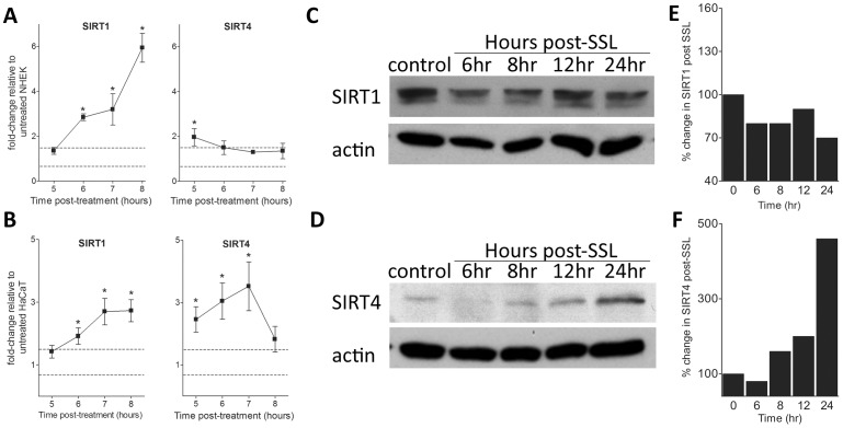
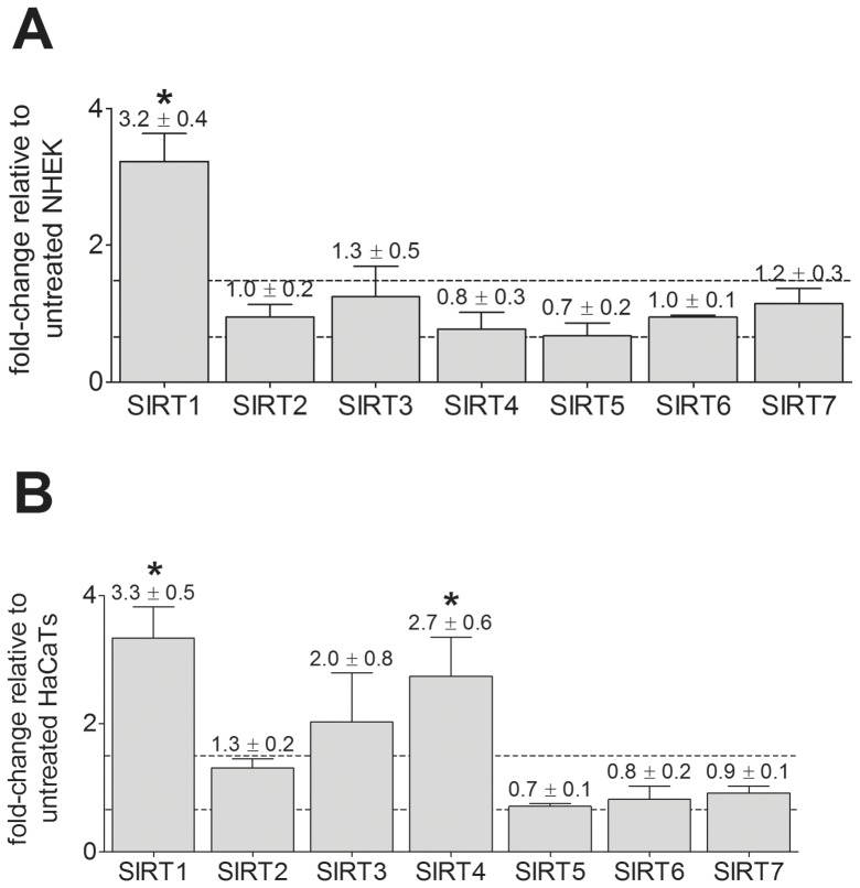
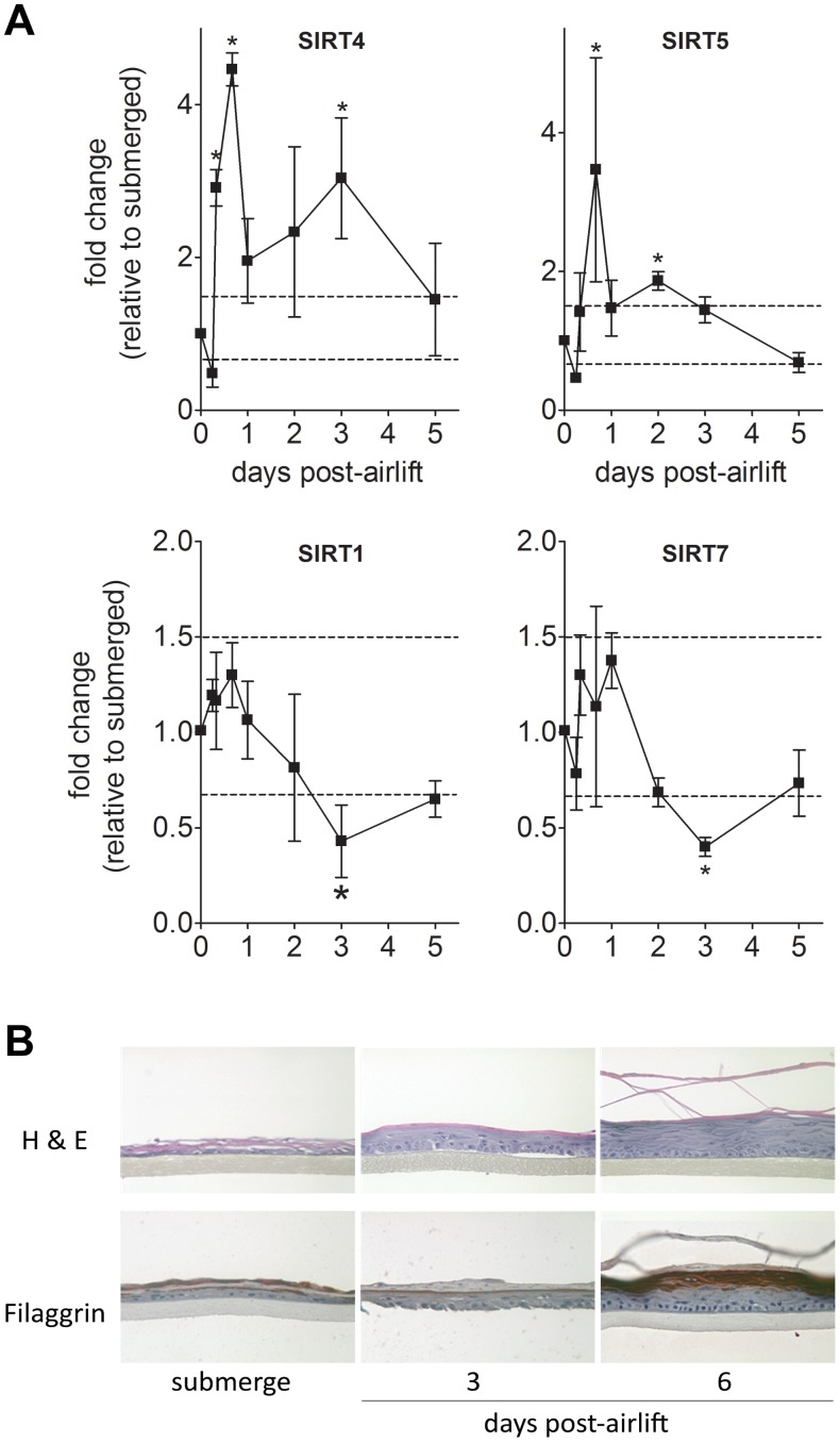
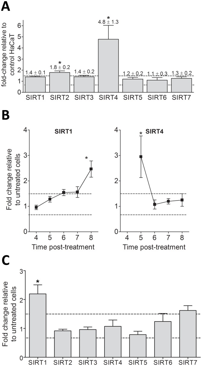
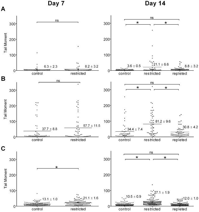
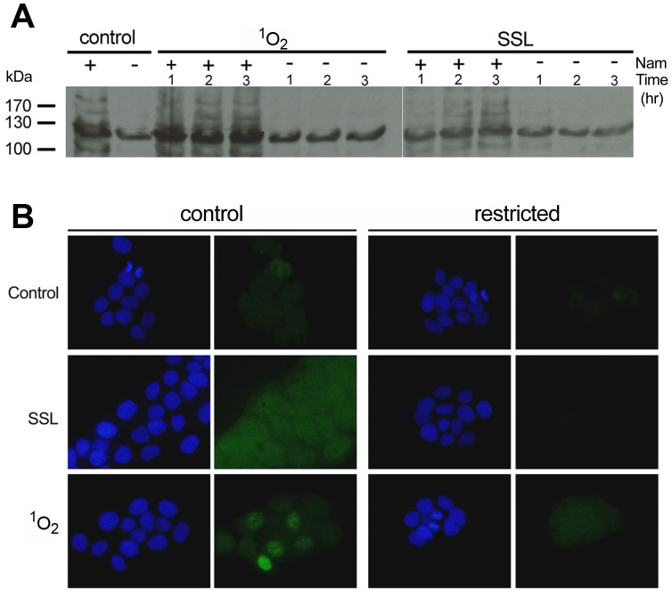
Sirtuins (SIRTs) and poly(ADP-ribose) polymerases (PARPs), NAD(+)-dependent enzymes, link cellular energy status with responses to environmental stresses. Skin is frequently exposed to the DNA damaging effects of UV irradiation, a known etiology in skin cancer. Thus, understanding the defense mechanisms in response to UV, including the role of SIRTs and PARPs, may be important in developing skin cancer prevention strategies. Here, we report expression of the seven SIRT family members in human skin. SIRTs gene expressions are progressively upregulated in A431 epidermoid carcinoma cells (SIRTs1 and 3), actinic keratoses (SIRTs 2, 3, 5, 6, and 7) and squamous cell carcinoma (SIRTs 1-7). Photodamage induces dynamic changes in SIRT expression with upregulation of both SIRT1 and SIRT4 mRNAs. Specific losses of SIRT proteins occur early after photodamage followed by accumulation later, especially for SIRT4. Niacin restriction, which decreases NAD(+), the sirtuin substrate, results in an increase in acetylated proteins, upregulation of SIRTs 2 and 4, increased inherent DNA damage, alterations in SIRT responses to photodamage, abrogation of PARP activation following photodamage, and increased sensitivity to photodamage that is completely reversed by repleting niacin. These data support the hypothesis that SIRTs and PARPs play important roles in resistance to photodamage and identify specific SIRTs that respond to photodamage and may be targets for skin cancer prevention.


Conflict of interest statement

Competing Interests: The authors have declared that no competing interests exist.

Figures

Figure 1
Figure 1. SIRT1 and SIRT4 gene and protein expression changes in NHEK and HaCaT cells following SSL treatment. (A–B)
Time course of SIRT1 and SIRT4 gene expression after SSL treatment was measured using qPCR. (A) NHEK cells (mean ± SEM, n = 2 independent experiments, each ran in triplicate). (B) HaCaT cells (mean ± SEM, n = 3 independent experiments, each ran in triplicate). Gene expression levels shown are expressed as fold-change relative to untreated cells, where all samples were normalized to GAPDH gene expression. *p<0.05 and a fold change >1.5 or <0.67 (dashed lines). (C) SIRT1 and (D) SIRT4 protein expression in NHEK cells was detected by Western blotting using the antibody described in Materials and Methods. To normalize for protein loading, blots were probed with an antibody against α-tubulin. (E) Percentage change in SIRT1 and (F) SIRT4 protein expression level relative to untreated NHEK after normalization to α-tubulin.
Figure 2
Figure 2. SIRT gene expression changes in keratinocytes following singlet oxygen stress.
SIRT gene expression at 5 hours after singlet oxygen stress treatment was measured using qPCR. (A) NHEK (mean ± SEM, n = 2 independent experiments with triplicate samples). (B) HaCaT cells (mean ± SEM, n = 3 independent experiments with triplicate samples). Gene expression levels shown are expressed as fold-change relative to untreated cells, where all samples were normalized to GAPDH gene expression. *p<0.05 and a fold change >1.5 or <0.67 (dashed lines).
Figure 3
Figure 3. SIRT mRNA expression changes during keratinocyte differentiation.
NHEK grown on a collagen-coated membrane were raised to the air-liquid interface (air-lift) to induce differentiation. (A) SIRT gene expression was measured by qPCR as a function of time. Expression levels shown are calculated as fold change relative to expression levels in submerged cultures (before airlift) after normalization to 18 S rRNA expression. Mean ± SEM, n = 4. *p<0.05 and a fold change >1.5 or <0.67 (dashed lines). (B) Hematoxylin and eosin (H&E) and filaggrin immunohistochemical staining of epidermal reconstructs reveals differentiation progress after airlift.
Figure 4
Figure 4. SIRT gene expression changes in HaCaT keratinocytes upon niacin restriction and photodamage.
SIRT gene expression profiles in HaCaT keratinocytes were measured using qPCR. (A) Niacin-restricted SIRT gene message levels shown are expressed as fold-change relative to niacin replete HaCaTs. (B) Time course of SIRT1 and SIRT4 mRNA expression after SSL treatment. (C) Time course of SIRT gene expression after singlet oxygen stress. Expression levels shown for B and C are calculated as fold-change relative to untreated niacin-restricted HaCaTs. All samples were normalized to GAPDH gene expression. Mean ± SEM, n = 3 independent experiments with triplicate samples. *p<0.05 and a fold change >1.5 or <0.67 (dashed lines).
Figure 5
Figure 5. Effect of photodamage on DNA integrity in niacin-restricted HaCaT keratinocytes.
DNA damage in HaCaT keratinocytes grown in the presence (control) or absence (restricted) of added niacin for 7 or 14 days was analyzed by alkaline comet assays. Dots represent the tail moment of single cells; mean tail moment is indicated by the lines shown for: (A) untreated, (B) SSL treated, and (C) singlet oxygen stress treated cells. ns, non significant difference; *, denotes significant differences, by unpaired Student’s t test, p<0.001.
Figure 6
Figure 6. Effect of niacin restriction on PARP activation by photodamage.
Polymer immunoblotting and immunocytochemistry of HaCaT keratinocytes grown for 14 days under control (+) or restricted (−) niacin (Nam) and exposed to photodamage. Cells treated with singlet oxygen stress (1O2) or solar simulated light (SSL) were analyzed by (A) Western blots using a PAR antibody and (B) immunostaining using a PAR antibody (green) and nuclear counterstaining with DAPI (blue).

Similar articles

Cited by

References

    1. Spronck JC, Kirkland JB (2002) Niacin deficiency increases spontaneous and etoposide-induced chromosomal instability in rat bone marrow cells in vivo. Mutat Res 508: 83–97. - PubMed
    1. Zhang JZ, Henning SM, Swendseid ME (1993) Poly(ADP-ribose) polymerase activity and DNA strand breaks are affected in tissues of niacin-deficient rats. J Nutr 123: 1349–1355. - PubMed
    1. Spronck JC, Nickerson JL, Kirkland JB (2007) Niacin Deficiency Alters p53 Expression and Impairs Etoposide-Induced Cell Cycle Arrest and Apoptosis in Rat Bone Marrow Cells. Nutr Cancer 57: 88–99. - PubMed
    1. Spronck JC, Bartleman AP, Boyonoski AC, Kirkland JB (2003) Chronic DNA damage and niacin deficiency enhance cell injury and cause unusual interactions in NAD and poly(ADP-ribose) metabolism in rat bone marrow. Nutr Cancer 45: 124–131. - PubMed
    1. Boyonoski AC, Gallacher LM, ApSimon MM, Jacobs RM, Shah GM, et al. (1999) Niacin deficiency increases the sensitivity of rats to the short and long term effects of ethylnitrosourea treatment. Mol Cell Biochem 193: 83–87. - PubMed

Publication types