. 2012;7(7):e42289.
doi: 10.1371/journal.pone.0042289. Epub 2012 Jul 31.

Vaccination with embryonic stem cells protects against lung cancer: is a broad-spectrum prophylactic vaccine against cancer possible?

Affiliations

Vaccination with embryonic stem cells protects against lung cancer: is a broad-spectrum prophylactic vaccine against cancer possible?

Kavitha Yaddanapudi et al. PLoS One. 2012.

Abstract

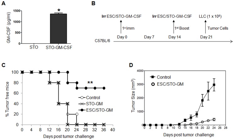
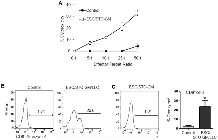
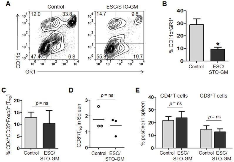
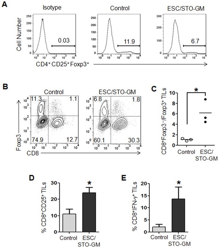
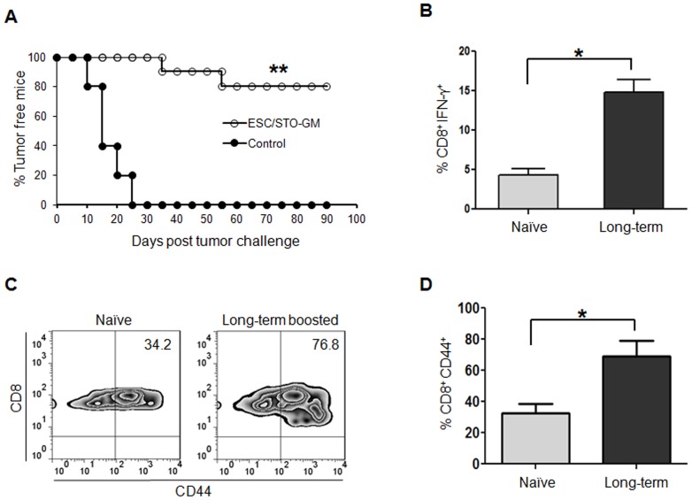
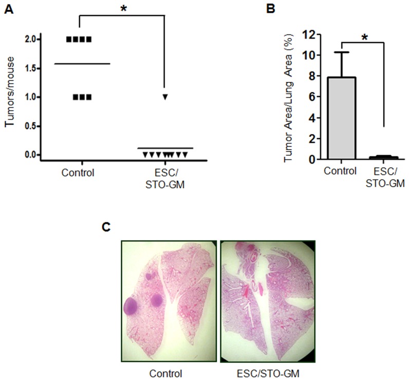
The antigenic similarity between tumors and embryos has been appreciated for many years and reflects the expression of embryonic gene products by cancer cells and/or cancer-initiating stem cells. Taking advantage of this similarity, we have tested a prophylactic lung cancer vaccine composed of allogeneic murine embryonic stem cells (ESC). Naïve C57BL/6 mice were vaccinated with ESC along with a source of granulocyte macrophage-colony stimulating factor (GM-CSF) in order to provide immunostimulatory adjuvant activity. Vaccinated mice were protected against subsequent challenge with implantable Lewis lung carcinoma (LLC). ESC-induced anti-tumor immunity was not due to a non-specific "allo-response" as vaccination with allogeneic murine embryonic fibroblasts did not protect against tumor outgrowth. Vaccine efficacy was associated with robust tumor-reactive primary and memory CD8(+) T effector responses, Th1 cytokine response, higher intratumoral CD8(+) T effector/CD4(+)CD25(+)Foxp3(+) T regulatory cell ratio, and reduced myeloid derived suppressor cells in the spleen. Prevention of tumorigenesis was found to require a CD8-mediated cytotoxic T lymphocyte (CTL) response because in vivo depletion of CD8(+) T lymphocytes completely abrogated the protective effect of vaccination. Importantly, this vaccination strategy also suppressed the development of lung cancer induced by the combination of carcinogen administration and chronic pulmonary inflammation. Further refinement of this novel vaccine strategy and identification of shared ESC/tumor antigens may lead to immunotherapeutic options for lung cancer patients and, perhaps more importantly, could represent a first step toward the development of prophylactic cancer vaccines.


Conflict of interest statement

Competing Interests: The authors have the following interests: The effects reported here are the subject of a pending U.S. Letters patent application on which Drs. Mitchell and Eaton are named as inventors, application number 60/782,672 (U.S. PTO), entitled “Methods and material for immunization against cancer”. It was filed March 15, 2007 (following first submission March 15, 2006). To the best of the authors' knowledge, there has been no office action on it and it remains an application. There are no other patents, products in development or marketed products to declare. This does not alter the authors' adherence to all the PLoS ONE policies on sharing data and materials, as detailed online in the guide for authors.

Figures

Figure 1
Figure 1. ESC vaccination prevents the outgrowth of an implanted lung adenocarcinoma.
(A) Bar graph showing GM-CSF expression in non-transduced and retrovirally transduced STO fibroblasts. Error bars represent mean ± SD. *, p<0.05; relative to non-transduced STO cells; t test. (B) Scheme of immunization. Male C57BL/6 mice were immunized twice (days 0 and 14) with HBSS (control), or irradiated 1×10⁶ ESC+irradiated 1×10⁶ GM-CSF-expressing STO murine embryonic fibroblasts (STO-GM) s.c. in the right flank. Seven days after boost, mice were challenged with 1×10⁵ Lewis lung carcinoma cells (LLC) s.c. in the left flank. (C) C57BL/6 mice (10/group) were immunized twice (days 0 and 14) with HBSS (control), or irradiated 1×10⁶ ESC+irradiated 1×10⁶ STO-GM, or irradiated 1×10⁶ STO-GM cells alone s.c. in the right flank prior to s.c. challenge with LLC on day 21. Tumor growth was monitored daily in all animals until sacrifice due to tumors exceeding 5% of body weight. The vaccinated tumor free mice remained so for up to 4 months later with no overt signs of distress or autoimmunity. Results are representative of three independent experiments. **, p<0.001; relative to control group; log-rank test. (D). Tumor growth was measured by calipers every 2nd or 3rd day and tumor volumes were plotted as indicated. The data represent the average tumor volumes of 10 mice/control group and 3 mice/ESC/STO-GM group and are representative of three independent experiments. Error bars represent mean ± SEM.
Figure 2
Figure 2. ESC vaccination elicits tumor cell-specific CD8-dependent cytotoxic response.
(A) C57BL/6 mice (5/group) were immunized twice (days 0 and 14) with HBSS (control) or irradiated 1×10⁶ ESC+irradiated 1×10⁶ STO-GM cells s.c. in the right flank. Ten days after boost, mice were euthanized and spleens were removed. Splenocytes from vaccinated and control mice were added to wells pre-seeded with LLC cells and co-cultured for an additional 16 hours with the indicated effector-to-target cell ratios, with loss of the latter monitored in an Acea electrical impedance reader. Results shown represent four independent wells for each effector-to-target ratio and error bars represent mean ± SD. (B–D) C57BL/6 mice were immunized twice (days 0 and 14) with HBSS (control) or irradiated 1×10⁶ ESC+irradiated 1×10⁶ STO-GM cells s.c. in the right flank. Seven days after the last immunization, mice were challenged with 1×10⁵ LLC cells s.c. in the left flank. 18–21 days after tumor challenge, mice were euthanized and spleens were removed. Additionally, one set of mice were vaccinated with ESC/STO-GM alone and not challenged with tumor. Splenocytes from vaccinated/tumor challenged, vaccinated/non-tumor challenged and control mice were washed, Fc receptors were blocked, and stained for surface expression of CD8 and intracellular expression of Granzyme B and analyzed by flow cytometry. (B, D) Percentage of CD8+ cells expressing granzyme B in splenocytes obtained from vaccinated/tumor challenged and control mice (6/group). (C) Percentage of CD8+ cells expressing granzyme B in splenocytes obtained from vaccinated/non-tumor challenged mice (6/group). Results are expressed as percentages of gated CD8+ splenocytes (*, p<0.05; relative to control group; t test). Error bars represent mean ± SD.
Figure 3
Figure 3. ESC vaccination induces tumor cell-specific, Th1-mediated cytokine response in CD8+ T cells.
C57BL/6 mice (6/group) were immunized twice (days 0 and 14) with HBSS (control) or irradiated 1×10⁶ ESC alone, or irradiated 1×10⁶ ESC+irradiated 1×10⁶ STO-GM, or irradiated 1×10⁶ STO-GM cells alone, s.c. in the right flank. Ten days after the boost, mice were euthanized and spleens were removed. Splenocytes from vaccinated and control mice were co-cultured with LLC lysate (50 µg/ml) for an additional 4 days. Effectors were harvested and stimulated for 4 hours with PMA (50 ng/ml) and ionomycin (500 ng/ml) in the presence of Brefeldin A (1 µl/ml). After restimulation, effectors were harvested, Fc receptors were blocked, and stained for surface expression of CD4, CD8 and intracellular expression of cytokines and analyzed by flow cytometry. (A, B) Dot plots showing TNF-α and IFN-γ expression in CD8+ cells in splenocyte cultures obtained from control and ESC/STO-GM vaccinated mice. Numbers in quadrants represent the percentages of each subpopulation. (C, D) Bar graphs showing percentages of CD8+TNF-α+, CD8+IFN-γ+, and CD8+IL2+ cells in splenocyte cultures derived from control, ESC alone, STO-GM alone and ESC/STO-GM vaccinated mice. Results are expressed as percentages of total cells. *, p<0.05; relative to control group; ANOVA. (E, F) Unstimulated spleen cells from vaccinated and control mice were directly treated with PMA/ionomycin/Brefeldin A and stained for intracellular cytokine expression. Two independent cell culture assays were performed with cells isolated from 6 mice per group; data from one representative assay is shown. Error bars represent mean ± SD.
Figure 4
Figure 4. ESC vaccination reduces myeloid-derived suppressor cells but does not alter T regulatory cells in the spleen.
C57BL/6 mice were immunized twice (days 0 and 14) with HBSS (control) or irradiated 1×10⁶ ESC+irradiated 1×10⁶ STO-GM cells s.c. in the right flank. Seven days after the last immunization, mice were challenged with 1×10⁵ LLC cells s.c. in the left flank. 18–21 days after tumor challenge, mice were euthanized and spleens were removed. Splenocytes from vaccinated and control mice were washed, Fc receptors were blocked, and stained for surface expression of different markers and analyzed by flow cytometry. (A) Dot plots showing percentages of splenic CD11b+GR1+ myeloid-derived suppressor cells (MDSCs) in control and ESC/STO-GM vaccinated mice. Numbers in quadrants represent the percentages of each subpopulation. (B) Bar graphs showing percentages of CD11b+GR1+ MDSCs in splenocytes obtained from control and ESC/STO-GM vaccinated mice. Results are expressed as percentages of total cells. *, p<0.05; relative to control group; t test. (C) Bar graphs showing percentages of CD4+CD25+Foxp3+ T regulatory cells (Treg) in splenocytes obtained from control and ESC/STO-GM vaccinated mice. Results are expressed as percentages of total cells. (D) The ratio of CD8+Foxp3 to CD4+CD25+Foxp3+ Treg cells was calculated and compared in splenocytes obtained from control and ESC/STO-GM vaccinated mice. (E) Bar graph showing percentages of CD4+ T and CD8+ T cells in splenocytes obtained from control and ESC/STO-GM vaccinated mice. Results are expressed as percentages of total cells. Three independent analyses were performed with cells isolated from 5 mice per group; data from one representative assay is shown. Error bars represent mean ± SD.
Figure 5
Figure 5. ESC vaccination increases the ratio of effector CD8+ T cells to Tregs in the tumor.
C57BL/6 mice were immunized twice (days 0 and 14) with HBSS (control) or irradiated 1×10⁶ ESC+irradiated 1×10⁶ STO-GM cells s.c. in the right flank. Seven days after boost, mice were challenged with 1×10⁵ Lewis lung carcinoma cells s.c. in the left flank. 18–21 days after tumor challenge, tumor-infiltrating cells were harvested from control and ESC/STO-GM vaccinated mice and analyzed by flow cytometry. (A) Histograms showing the percentages of CD4+CD25+Foxp3+ Tregs in CD45.2+ tumor infiltrating cells obtained from control and ESC/STO-GM vaccinated mice. (B) Dot plots showing the percentages of CD8+ and Foxp3+ sub-populations in CD45.2+ tumor infiltrating cells. (C) Bar graph showing the ratio of CD8+ Foxp3 to CD8Foxp3+ cells in 1 of 2 representative experiments with 3 independently analyzed mice/group. *, p<0.05; relative to control group; t test. (D, E) ESC vaccination increases the frequency of functional CD8+ T cells in tumors. (D) Bar graph showing the percentages of CD25+CD8+ in CD45.2+ tumor infiltrating cells obtained from control and ESC/STO-GM vaccinated mice. (E) Tumor infiltrating cells were restimulated with PMA and ionomycin and analyzed for the expression of intracellular IFN-γ. Bar graphs showing percentages of CD8+IFN-γ+ in tumor infiltrating cells from control and ESC/STO-GM vaccinated mice. Results are expressed as percentages of total cells. The data represent results from 2 independent experiments with 3 mice/group. *, p<0.05; relative to control group; t test. Error bars represent mean ± SD.
Figure 6
Figure 6. ESC vaccination-induced CD8+ T-effector responses are maintained in long-term surviving animals.
Long-term surviving mice from the ESC/STO-GM group were re-challenged s.c with 1×10⁵ LLC cells 60 days after initial tumor inoculation. (A) Tumor growth was monitored daily in all animals until sacrifice due to tumors exceeding 5% of body weight. Results represent a summation of two independent experiments with 10 mice/group. **, p<0.001; relative to naïve control; log-rank test. (B–D) Spleens were isolated from vaccinated survivors and control naïve mice 10 days after the tumor injection. (B) Splenocytes were harvested and stimulated for 4 hours with PMA (50 ng/ml) and ionomycin (500 ng/ml) in the presence of Brefeldin A (1 µl/ml). After restimulation, cells were harvested, Fc receptors were blocked, and stained for intracellular expression of cytokines, and analyzed by flow cytometry. Bar graphs showing percentage of intracellular IFN-γ expression in CD8+ cells (*, p<0.05; relative to naïve control; t test). Results are expressed as percentages of total cells. (C) Dot plots showing the percentages of CD44+ and CD8+ cells in splenocytes derived from long-term surviving ESC vaccinated and control naïve mice. Numbers in the quadrants represent percentages of each subpopulation. (D) Bar graph showing the percentages of CD44+CD8+ cells (*, p<0.05; relative to naïve control; t test). Results are expressed as percentages of total cells. Data for B–D are representative of two independent experiments with 4 mice/group. Error bars represent mean ± SD.
Figure 7
Figure 7. ESC vaccination suppresses 3-methylcholanthrene initiated, butylated hydroxytoluene promoted lung carcinogenesis.
(A) Eighteen weeks after administration of the single dose of 3-methylcholanthrene followed by repetitive doses of BHT, lungs from euthanized Balb/c control and ESC/STO-GM vaccinated mice (8/group) were resected and inflated at a pressure of 15 cm with 4% buffered formalin. Surface tumors were enumerated by inspection under 5× magnification. *, p<0.05; relative to control group; t test. (B) The percentage of total lung area taken up by adenocarcinomas was quantified from measurements on H&E stained serial sections of lungs from three animals in each group (sections examined were 100 µm apart). *, p<0.05; relative to control group; t test. Error bars represent mean ± SD. (C) Representative H&E stained sections from control or ESC/STO-GM vaccinated mice (8/group) were photographed under low power (5×).

Similar articles

Cited by

References

    1. Triolo VA (1965) Nineteenth Century Foundations of Cancer Research Advances in Tumor Pathology, Nomenclature, and Theories of Oncogenesis. Cancer Res 25: 75–106. - PubMed
    1. Al-Hajj M, Clarke MF (2004) Self-renewal and solid tumor stem cells. Oncogene 23: 7274–7282. - PubMed
    1. Stonehill EH, Bendich A (1970) Retrogenetic expression: the reappearance of embryonal antigens in cancer cells. Nature 228: 370–372. - PubMed
    1. Baldwin RW, Glaves D, Vose BM (1972) Embryonic antigen expression in chemically induced rat hepatomas and sarcomas. Int J Cancer 10: 233–243. - PubMed
    1. Baldwin RW, Glaves D, Pimm MV, Vose BM (1972) Tumour specific and embryonic antigen expression of chemically induced rat tumours. Ann Inst Pasteur (Paris) 122: 715–728. - PubMed

Publication types

MeSH terms