

doi: 10.1021/cn3000058.
Epub 2012 Feb 17.
BODIPY-based molecular probe for imaging of cerebral β-amyloid plaques
Affiliations
- PMID: 22860198
- PMCID: PMC3369805
- DOI: 10.1021/cn3000058
Item in Clipboard
BODIPY-based molecular probe for imaging of cerebral β-amyloid plaques
ACS Chem Neurosci.
.
Abstract
We designed and synthesized a BODIPY-based probe (BAP-1) for the imaging of β-amyloid plaques in the brain. In binding experiments in vitro, BAP-1 showed excellent affinity for synthetic Aβ aggregates. β-Amyloid plaques in Tg2576 mouse brain were clearly visualized with BAP-1. In addition, the labeling of β-amyloid plaques was demonstrated in vivo in Tg2576 mice. These results suggest BAP-1 to be a useful fluorescent probe for the optical imaging of cerebral β-amyloid plaques in patients with Alzheimer's disease.
Keywords: Alzheimer’s disease; BODIPY; optical imaging; β-amyloid plaque.
Figures
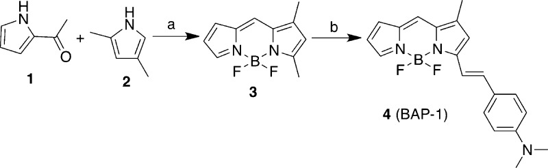
Reagents: (a) CHCl3, POCl3, BF3OEt2, Et3N; (b) toluene, 4-dimethylaminobenzaldehyde, piperidine, and
AcOH.

Fluorescence intensity of BAP-1 upon interaction with Aβ42
aggregates and BSA.
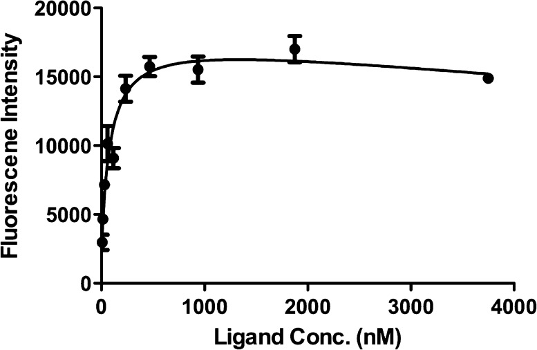
Plot of the fluorescence
intensity (Em = 673 nm) as a function
of the concentration of BAP-1 in the presence of Aβ42 aggregates
(2.2 μM) in solutions.
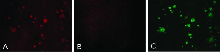
Neuropathological staining
of BAP-1 in a 10-μm section from
a Tg2576 mouse brain (A) and a wild-type mouse brain (B). Labeled
plaques were confirmed by staining of the adjacent section with thioflavin-S
(C).
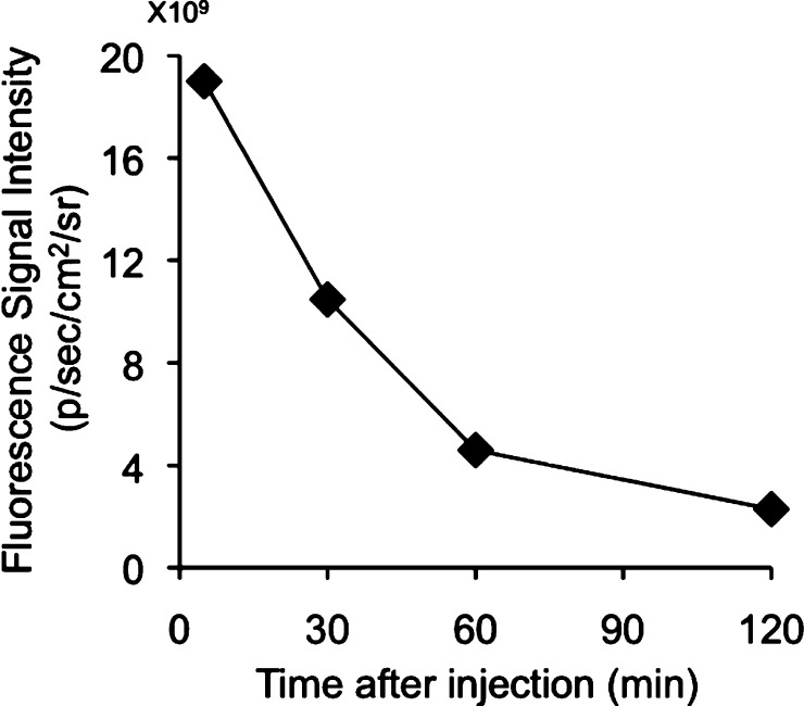
Fluorescence intensity after injection of BAP-1 into ddY
mice with
an IVIS spectrum.
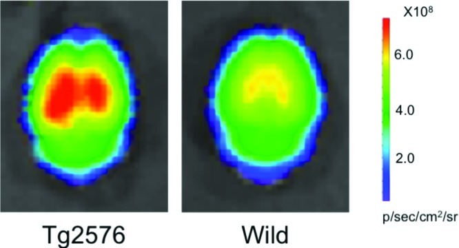
Comparison of the fluorescence intensity in the brain
after the
injection of BAP-1 into a Tg2576 mouse (A) and wild-type mouse (B).
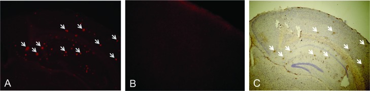
Ex vivo
fluorescence observation of brain sections from a Tg2576
mouse (A) and wild-type mouse (B) after an injection of BAP-1. The
presence of Aβ plaques in the section from the Tg2576 mouse
was confirmed with immunohistochemical staining using a monoclonal
Aβ antibody (C). Arrows show Aβ plaques stained by both
BAP-1 (A) and immunohistochemical labeling (C).
References
-
- Klunk W. E. (1998) Biological markers of Alzheimer’s disease. Neurobiol. Aging 19, 145–147. - PubMed
-
- Selkoe D. J. (2000) Imaging Alzheimer’s amyloid. Nat. Biotechnol. 18, 823–824. - PubMed
-
- Ono M.; Wilson A.; Nobrega J.; Westaway D.; Verhoeff P.; Zhuang Z. P.; Kung M. P.; Kung H. F. (2003) 11C-labeled stilbene derivatives as Aβ-aggregate-specific PET imaging agents for Alzheimer’s disease. Nucl. Med. Biol. 30, 565–571. - PubMed
-
- Verhoeff N. P.; Wilson A. A.; Takeshita S.; Trop L.; Hussey D.; Singh K.; Kung H. F.; Kung M. P.; Houle S. (2004) In-vivo imaging of Alzheimer disease β-amyloid with [11C]SB-13 PET. Am. J. Geriatr. Psychiatry 12, 584–595. - PubMed
-
- Mathis C. A.; Wang Y.; Holt D. P.; Huang G. F.; Debnath M. L.; Klunk W. E. (2003) Synthesis and evaluation of 11C-labeled 6-substituted 2-arylbenzothiazoles as amyloid imaging agents. J. Med. Chem. 46, 2740–2754. - PubMed
Publication types
MeSH terms
Substances
LinkOut - more resources
Full Text Sources
Other Literature Sources
Molecular Biology Databases
