Review
. 2012 Sep;169(3):205-12.
doi: 10.1111/j.1365-2249.2012.04602.x.

Lymph node dissection--understanding the immunological function of lymph nodes

Affiliations
Review

Lymph node dissection--understanding the immunological function of lymph nodes

M Buettner et al. Clin Exp Immunol. 2012 Sep.

Abstract

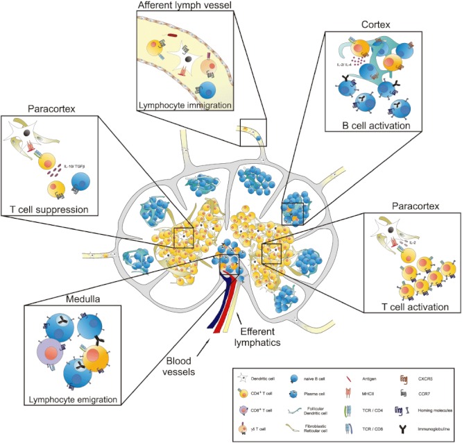
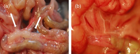
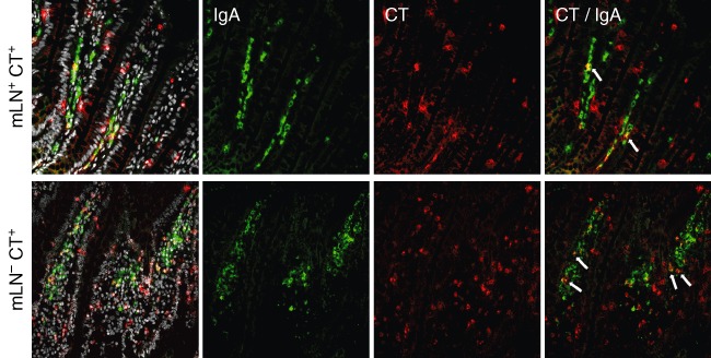
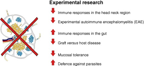
Lymph nodes (LN) are one of the important sites in the body where immune responses to pathogenic antigens are initiated. This immunological function induced by cells within the LN is an extensive area of research. To clarify the general function of LN, to identify cell populations within the lymphatic system and to describe the regeneration of the lymph vessels, the experimental surgical technique of LN dissection has been established in various animal models. In this review different research areas in which LN dissection is used as an experimental tool will be highlighted. These include regeneration studies, immunological analysis and studies with clinical questions. LN were dissected in order to analyse the different cell subsets of the incoming lymph in detail. Furthermore, LN were identified as the place where the induction of an antigen-specific response occurs and, more significantly, where this immune response is regulated. During bacterial infection LN, as a filter of the lymph system, play a life-saving role. In addition, LN are essential for the induction of tolerance against harmless antigens, because tolerance could not be induced in LN-resected animals. Thus, the technique of LN dissection is an excellent and simple method to identify the important role of LN in immune responses, tolerance and infection.


Figures

Fig. 1
Fig. 1
Function of the lymph nodes (LN). LN are the site where immune reactions or suppression takes place. Cells of the afferent or efferent lymphatics are collected via LN dissection and the cells can be analysed. As well as different dendritic cell (DC) populations, different lymphocyte subpopulations such as CD4+ T cells or γδ T cells are also detected. Within the paracortex of the lymph node, DC present antigens to T cells which proliferate and differentiate into effector or helper T cells after recognizing the specific antigen whereby an immune response is initiated. T helper cells migrate into the cortex to assist B cells to proliferate and differentiate into effector or plasma cells. All lymphocytes, including naive or effector cells, migrate to the medulla to leave the LN via the efferent lymphatics or the blood system. Conversely, oral tolerance is activated if regulatory T cells are activated via DC, which prevent immune response induction. Without the LN all these functions are reduced or lacking.
Fig. 2
Fig. 2
The gut system with and without mesenteric lymph nodes (mLN). Mice were anaesthetized and the abdomen was opened. The gut was taken out and the mLN were seen (a, mLN indicated by arrows). The mLN were removed carefully. Afterwards the gut was replaced in the abdomen and the abdomen was closed. After 4 weeks animals were studied by feeding oil, which is transported via the lymphatics, to check for newly connected pseudo-afferent lymphatics (b).
Fig. 3
Fig. 3
The number of antigen-specific immunoglobulin (Ig)A+ cells in the lamina propria is increased after mesenteric lymph node (mLN) resection. Immunofluorescence staining of the lamina propria of the gut in mLN-bearing and mLN-resected rats, which had been treated with cholera toxin (CT), was carried out with antibodies against IgA (green) and CT (red); 4',6-diamidino-2-phenylindole (DAPI) was used to visualize all cells. CT-specific IgA+ cells (arrows) are seen in both groups, but to a greater extent in mLN-resected animals.
Fig. 4
Fig. 4
The role of lymph nodes (LN) in research. LN are unique organs distributed all over the body. They have to filter the lymph for incoming antigens from the draining area to defend the body. Removing the LN leads to various different responses in the body. For example, the immune response in the gut system is enhanced after LN dissection, whereas the immune responses in the head–neck region are diminished.

References

    1. Pabst R. Plasticity and heterogeneity of lymphoid organs. What are the criteria to call a lymphoid organ primary, secondary or tertiary? Immunol Lett. 2007;112:1–8. - PubMed
    1. Sainte-Marie G. The lymph node revisited: development, morphology, functioning, and role in triggering primary immune responses. Anat Rec (Hoboken) 2010;293:320–37. - PubMed
    1. Ahrendt M, Hammerschmidt SI, Pabst O, Pabst R, Bode U. Stromal cells confer lymph node-specific properties by shaping a unique microenvironment influencing local immune responses. J Immunol. 2008;181:1898–907. - PubMed
    1. Hammerschmidt SI, Ahrendt M, Bode U, et al. Stromal mesenteric lymph node cells are essential for the generation of gut-homing T cells in vivo. J Exp Med. 2008;205:2483–90. - PMC - PubMed
    1. Magnusson FC, Liblau RS, von Boehmer H, et al. Direct presentation of antigen by lymph node stromal cells protects against CD8 T-cell-mediated intestinal autoimmunity. Gastroenterology. 2008;134:1028–37. - PubMed

Publication types

MeSH terms

Substances