. 2013;22(1):87-97.
doi: 10.3727/096368912X653174. Epub 2012 Aug 2.

¹⁹F MRI tracer preserves in vitro and in vivo properties of hematopoietic stem cells

Affiliations

¹⁹F MRI tracer preserves in vitro and in vivo properties of hematopoietic stem cells

Brooke M Helfer et al. Cell Transplant. 2013.

Abstract

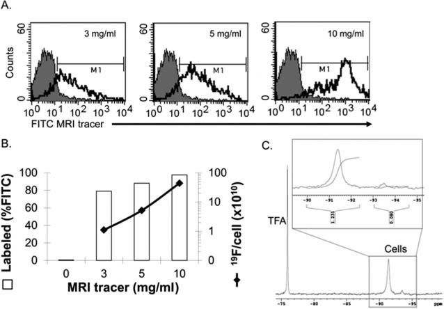
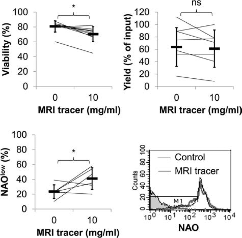
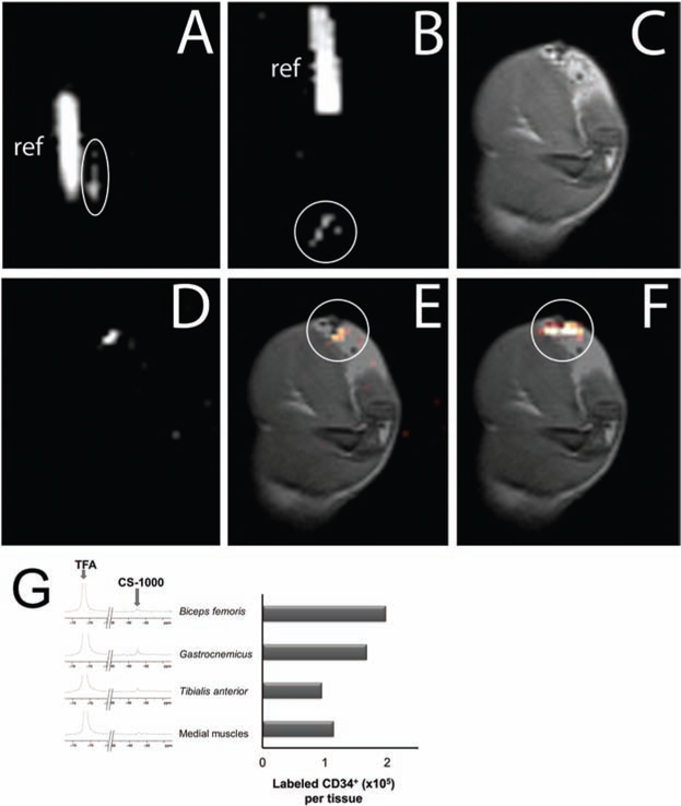
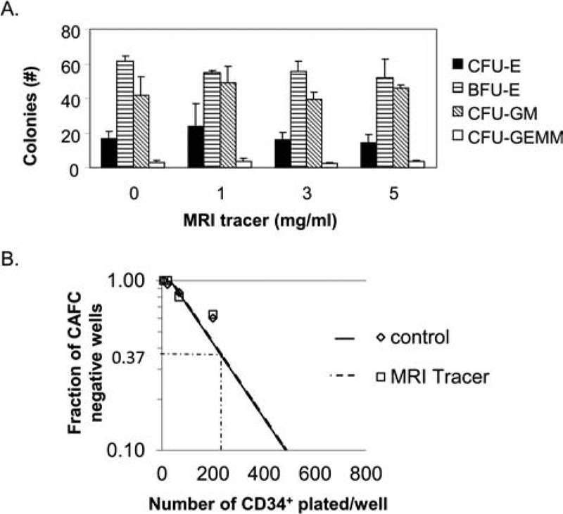
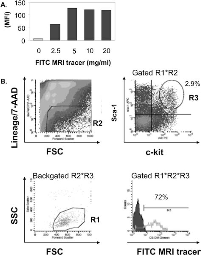
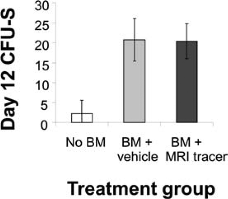
Hematopoietic stem cells (HSCs) have numerous therapeutic applications including immune reconstitution, enzyme replacement, regenerative medicine, and immunomodulation. The trafficking and persistence of these cells after administration is a fundamental question for future therapeutic applications of HSCs. Here, we describe the safe and efficacious labeling of human CD34(+) HSCs with a novel, self-delivering perfluorocarbon ¹⁹F magnetic resonance imaging (MRI) tracer, which has recently been authorized for use in a clinical trial to track therapeutic cells. While various imaging contrast agents have been used to track cellular therapeutics, the impact of this MRI tracer on HSC function has not previously been studied. Both human CD34(+) and murine bone marrow (BM) HSCs were effectively labeled with the MRI tracer, with only a slight reduction in viability, relative to mock-labeled cells. In a pilot study, ¹⁹F MRI enabled the rapid evaluation of HSC delivery/retention following administration into a rat thigh muscle, revealing the dispersal of HSCs after injection, but not after surgical implantation. To investigate effects on cell functionality, labeled and unlabeled human HSCs were tested in in vitro colony forming unit (CFU) assays, which resulted in equal numbers of total CFU as well as individual CFU types, indicating that labeling did not alter multipotency. Cobblestone assay forming cell precursor frequency was also unaffected, providing additional evidence that stem cell function was preserved after labeling. In vivo tests of multipotency and reconstitution studies in mice with murine BM containing labeled HSCs resulted in normal development of CFU in the spleen, compared to unlabeled cells, and reconstitution of both lymphoid and myeloid compartments. The lack of interference in these complex biological processes provides strong evidence that the function and therapeutic potential of the HSCs are likely maintained after labeling. These data support the safety and efficacy of the MRI tracer for clinical tracking of human stem cells.


Figures

Figure 1.
Figure 1.
Labeling efficiency of CD34+ hematopoietic stem cells (HSCs) with 19F MRI tracer. (A) Human bone marrow (BM) CD34+ cells were incubated with increasing doses of fluorescein isothiocyanate-magnetic resonance imaging (FITC-MRI) tracer to optimize labeling efficiency by flow cytometry. Autofluorescence in control cells is represented by the gray-filled histograms and the green fluorescence in MRI tracer labeled cells is indicated by the unfilled histograms (one of three experiments with similar results). (B) Average labeling efficiency versus increasing dose of MRI tracer. Left axis, open bars: percentage of the CD34+ cell population containing label as measured by flow cytometry fluorescent signal above background (denoted by M1 in A); right axis, solid line, perfluorocarbon (PFC) label uptake measured by 19F nuclear magnetic resonance (NMR). (C) Representative NMR spectra of 10 mg/ml MRI tracer labeled CD34+ cells corresponding to a 4.4 × 10¹¹ 19F atoms/cell labeling efficiency (one of three experiments with similar results). An inset depicts a typical integration curve, with the peak at −91 ppm used for labeling efficiency calculations.
Figure 2.
Figure 2.
CD34+ cellular viability is modestly reduced with MRI tracer. Results represent the paired analysis of repeated experiments assessing viability by Trypan blue, apoptosis by nonyl acridine orange (NAO) expression and yields after CD34+ cells were cultured with and without MRI tracer (10 mg/ml). Averages are indicated in heavy horizontal bars for viability (n = 10), NAO expression (n = 6), and yields (n = 7). Histograms of flow cytometric analysis of NAO staining in MRI tracer-labeled and unlabeled cells as well as unstained cells in shaded histogram. Cells undergoing apoptosis (NAOlow) are detected by a decrease in fluorescence (region defined by M1).
Figure 3.
Figure 3.
Imaging delivery of PFC-labeled human CD34+ cells in vivo with 19F/1H MRI. (A, B) In vitro 19F MRI of labeled CD34+ cells prior to administration: 15 × 106 cells in 1× PBS suspension for injection (A) and 14 × 106 CD34+ cells in a Matrigel support for implantation (B) (n = 2). Both labeled cells (circled) and reference tubes (“ref”) are in the field of view. In vitro images employ nonlinear brightness contrast adjustments to distinguish the cell signal from background and reference signal from saturation simultaneously. (C–F) In vivo MRI approximately 2 h after surgical implantation of the cell-laden Matrigel plug. (C) Proton image of the rat thigh anatomy with the hydrogel implant clearly visible as a hypointense region at the top right of the image. (D) 19F image (100-min acquisition) of the same field of view. (E) Coregistered 1H and 19F image overlay (C, D), with the 19F rendered in a “hot-iron” color scale (circled). Image quantification confirmed approximately 14 × 106 ± 2 × 10⁶ cells remain in the region of implant. (F) Overlay of postsacrifice, 19F scan of implanted cells (circled) on 1H anatomical image. (G) Ex vivo 19F NMR spectroscopy was conducted with dissected thigh muscles to evaluate the delivery of labeled CD34+ suspension cells injected using a syringe. Analyzed muscles included the site of injection (biceps femoris), muscles proximal to the injection site (gastrocnemius and tibialis anterior), and a set muscles distal to the injection site (“medial muscles” including portions of the semitendenous, gracilis, semimebranous, and adductor magnus). Spectra are depicted beside a chart of the quantification of transplanted cell numbers in each tissue. TFA, trifluoroacetic acid; CS-1000, Cell Sense tracer.
Figure 4.
Figure 4.
MRI tracer preserves differentiation and self-renewal of human CD34+ cells in vitro. (A) Human bone marrow CD34+ HSCs were incubated with or without increasing amounts of the MRI tracer for 4 h prior to seeding in methocellulose in triplicate for colony-forming unit (CFU) assays. Results show the average CFU per 1,000 progenitor cells/dish, and error bars indicate standard deviations. One of two independent studies with identical findings is depicted. (B) Limiting dilution of human bone marrow CD34+ HSCs (unlabeled and MRI tracer-labeled, 10 mg/ml), 20 replicate wells per condition, were cultured with stromal cells in the presence of serum, interleukin (IL)-3, and granulocyte macrophage-colony-stimulating factor (GM-CSF) for 5 weeks and then observed for the formation of cobblestone areas (cobblestone area forming cell, CAFC, assay). The frequency of negative wells plotted against the number of cells added per culture provides a graphical depiction of the Poisson equation to calculate precursor frequency in the treated and untreated cells. CFU-E, CFU-erythroid; BFU-E, blast forming unit-erythroid; CFU-GM, CFU-granulocyte, macrophage; CFU-GEMM, CFU-granulocyte, erythroid, macrophage, megakaryocyte.
Figure 5.
Figure 5.
MRI tracer labels murine bone marrow HSCs. (A) Increasing doses of FITC-MRI tracer was incubated with murine bone marrow mononuclear cells and then evaluated by flow cytometry. (B) Labeling of murine HSCs present in bone marrow was confirmed by flow cytometry. Gating strategy for enumeration of labeled HSCs is depicted and included selection of live cells by forward scatter (FSC) and side scatter (SSC) (R1), exclusion of dead 7-aminoactinomycin D (7-AAD)+ and peridinin-chlorophyll-protein complex (PerCP)-labeled lineage (lin)+ cells in the FL-3 channel (R2), and gating double-positive c-kit+ stem cell antigen (Sca)-1+ BM cells in FL-2 and FL-4 channels, respectively (R3), to examine live, 7-AAD lin Sca-1+ c-kit+ cells for the presence of FITC-MRI tracer (detected in FL-1 channel), lower right plot. One of three independent experiments with similar results is shown. Backgated R2*R3 scatter plot is provided to verify selection of the correct HSC population in lower left plot. MFI, mean fluorescence intensity.
Figure 6.
Figure 6.
HSC activity is preserved in vivo with the addition of MRI tracer. Myeloablated murine hosts were transplanted with labeled or unlabeled syngeneic bone marrow, with untransplanted mice serving as a negative control (n = 8 per cohort). On day 12 (day 11 for untransplanted recipients), spleens were harvested and CFU-S enumerated. Results represent the average CFU-S per animal. Significantly fewer numbers of colonies were measured in untransplanted recipients compared to labeled or unlabeled transplanted recipients (p < 0.0001 for each comparison). No significant differences in colony number were found between labeled and unlabeled cells when analyzed by unpaired t test (with or without Bonferroni correction).

References

    1. Ahrens ET; Flores R; Xu HY; Morel PA In vivo imaging platform for tracking immunotherapeutic cells. Nat. Biotechnol 23(8):983–987; 2005. - PubMed
    1. Arbab AS; Yocum GT; Kalish H; Jordan EK; Anderson SA; Khakoo AY; Read EJ; Frank JA Efficient magnetic cell labeling with protamine sulfate complexed to ferumoxides for cellular MRI. Blood 104(4):1217–1223; 2004. - PubMed
    1. Arbab AS; Yocum GT; Rad AM; Khakoo AY; Fellowes V; Read EJ; Frank JA Labeling of cells with ferumoxides-protamine sulfate complexes does not inhibit function or differentiation capacity of hematopoietic or mesenchymal stem cells. NMR Biomed. 18(8):553–559; 2005. - PubMed
    1. Boehm-Sturm P; Mengler L; Wecker S; Hoehn M; Kallur T In vivo tracking of human neural stem cells with 19F magnetic resonance imaging. PLoS One 6(12): e29040; 2011. - PMC - PubMed
    1. Bonetto F; Srinivas M; Heerschap A; Mailliard R; Ahrens ET; Figdor CG; de Vries IJ A novel (19)F agent for detection and quantification of human dendritic cells using magnetic resonance imaging. Int. J. Cancer 129(2):365–373; 2011. - PMC - PubMed

Publication types

LinkOut - more resources