

Comparison of cone-beam computed tomography incidental findings between patients with moderate/severe obstructive sleep apnea and mild obstructive sleep apnea/healthy patients
- PMID: 22862979
- PMCID: PMC3428237
- DOI: 10.1016/j.oooo.2012.03.014
Comparison of cone-beam computed tomography incidental findings between patients with moderate/severe obstructive sleep apnea and mild obstructive sleep apnea/healthy patients
Abstract
Objective: The objective of this study was to compare the incidental radiographic findings in the maxillofacial structures and the pharyngeal airway between subjects with moderate/severe obstructive sleep apnea (OSA) and mild OSA/healthy subjects using cone-beam computed tomography (CBCT) scans.
Study design: A total of 53 subjects with moderate/severe OSA (with a Respiratory Disturbance Index [RDI] ≥ 15 events/h) and 33 mild OSA/healthy subjects (RDI < 15), based on ambulatory somnographic assessment, were recruited. Supine CBCTs were taken and sent for radiological report. The incidental findings were compared between the 2 groups.
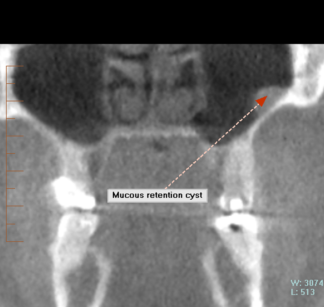
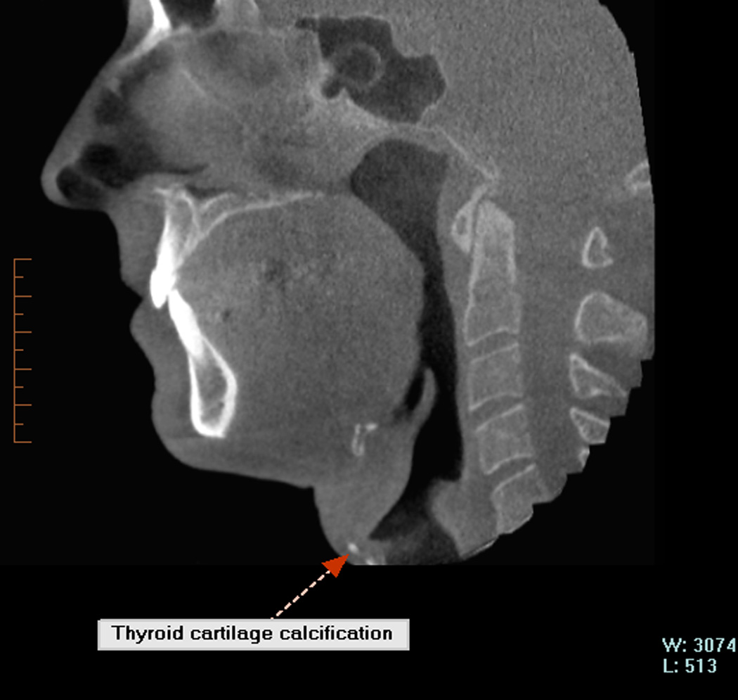
Results: Moderate/severe subjects had larger prevalence of conchae bullosa, hypertrophic turbinates, hypertrophic tonsils, elongated or posteriorly placed soft palate, narrower airway, enlarged tongue, and focal calcifications, although no significant differences were found.
Conclusions: CBCT is useful in identifying maxillofacial and airway anomalies that could interfere with normal breathing; however, no significant difference was found in prevalence of incidental findings between subjects with moderate/severe OSA and mild OSA/healthy subjects. Further studies are necessary to generalize our results.
Copyright © 2012 Elsevier Inc. All rights reserved.
Conflict of interest statement
The authors declare that they have no conflict of interest.
Figures


References
-
- Hatcher DC, Dial C, Mayorga C. Cone beam CT for pre-surgical assessment of implant sites. J Calif Dent Assoc. 2003;31:825–833. - PubMed
-
- Tyndall DA, Rathore S. Cone-beam CT diagnostic applications: caries, periodontal bone assessment, and endodontic applications. Dent Clin North Am. 2008 Oct;52(4):825–841. - PubMed
-
- Mah J, Enciso R, Jorgensen M. Management of impacted cuspids using 3-D volumetric imaging. J Calif Dent Assoc. 2003;31:835–841. - PubMed
-
- Erickson M, Caruso JM, Leggitt L. Newtom QR-DVT 9000 imaging used to confirm a clinical diagnosis of iatrogenic mandibular nerve paresthesia. J Calif Dent Assoc. 2003;31:843–845. - PubMed
-
- Bouquet A, Coudert JL, Bourgeois D, Mazoyer JF, Bossard D. Contributions of reformatted computed tomography and panoramic radiography in the localization of third molars relative to the maxillary sinus. Oral Surg Oral Med Oral Pathol Oral Radiol Endod. 2004;98:342–347. - PubMed
Publication types
MeSH terms
Grants and funding
LinkOut - more resources
Full Text Sources
