. 2012 Aug 3;150(3):481-94.
doi: 10.1016/j.cell.2012.07.012.

TPP1 OB-fold domain controls telomere maintenance by recruiting telomerase to chromosome ends

Affiliations

TPP1 OB-fold domain controls telomere maintenance by recruiting telomerase to chromosome ends

Franklin L Zhong et al. Cell. .

Abstract

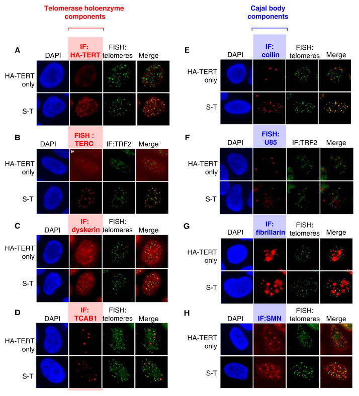
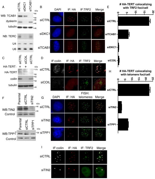
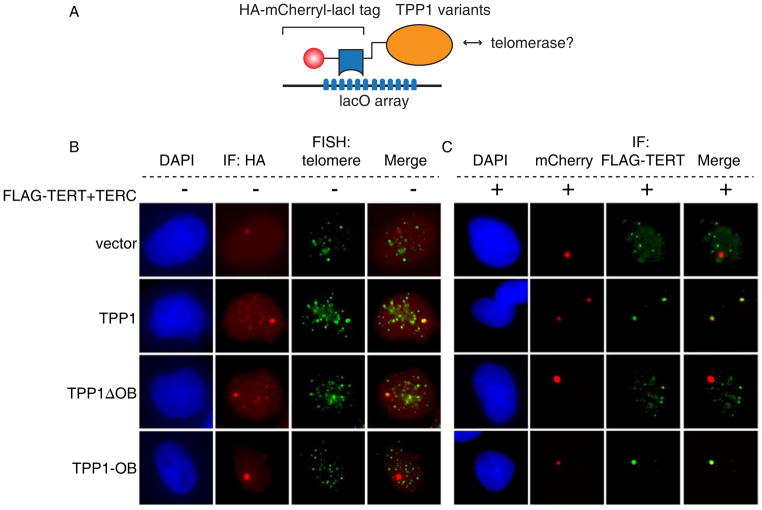
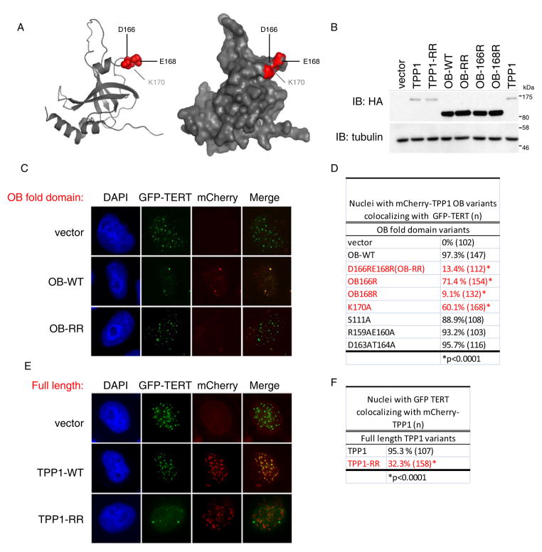
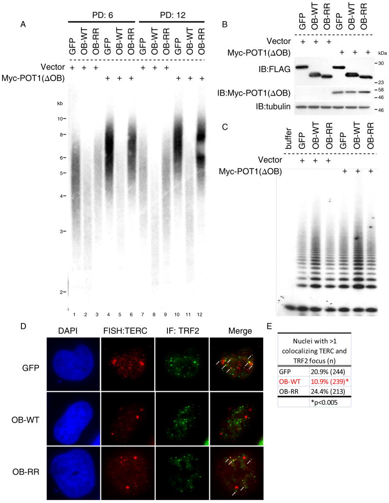
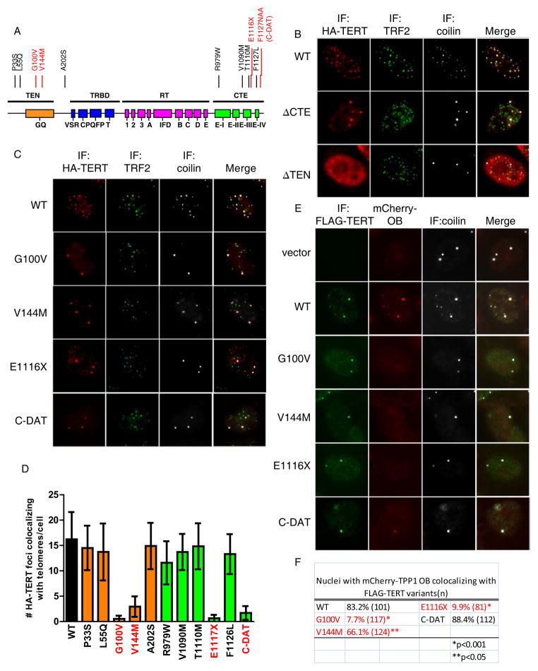
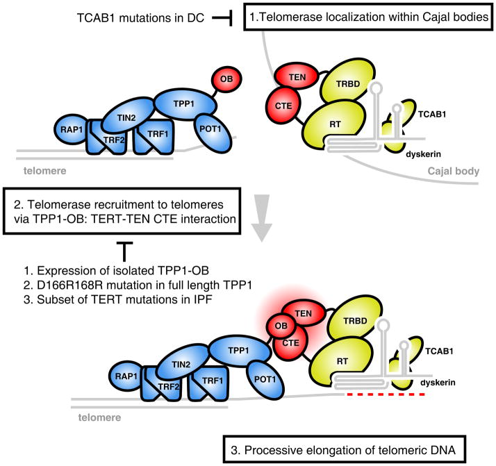
Telomere synthesis in cancer cells and stem cells involves trafficking of telomerase to Cajal bodies, and telomerase is thought to be recruited to telomeres through interactions with telomere-binding proteins. Here, we show that the OB-fold domain of the telomere-binding protein TPP1 recruits telomerase to telomeres through an association with the telomerase reverse transcriptase TERT. When tethered away from telomeres and other telomere-binding proteins, the TPP1 OB-fold domain is sufficient to recruit telomerase to a heterologous chromatin locus. Expression of a minimal TPP1 OB-fold inhibits telomere maintenance by blocking access of telomerase to its cognate binding site at telomeres. We identify amino acids required for the TPP1-telomerase interaction, including specific loop residues within the TPP1 OB-fold domain and individual residues within TERT, some of which are mutated in a subset of pulmonary fibrosis patients. These data define a potential interface for telomerase-TPP1 interaction required for telomere maintenance and implicate defective telomerase recruitment in telomerase-related disease.


Figures

Figure 1
Figure 1. Telomerase holoenzyme components at telomeres and formation of neo-Cajal bodies in S-T cells
HeLa cells transduced with an HA-TERT retrovirus were transfected with a TERC expression plasmid (S-T, super-telomerase) or empty vector (HA-TERT only). (A–D) Immunofluorescence or FISH for telomerase components and telomeres using: (A) anti-HA antibody (red), DNA FISH for telomeres (green); (B) RNA FISH for TERC (red), anti-TRF2 antibody (green); (C) anti-dyskerin antibody (red), DNA FISH for telomeres (green); (D) anti-TCAB1 antibodies (red), DNA FISH for telomeres (green). (E–H) Immunofluorescence or FISH for Cajal body components and telomeres using: (E) anti-coilin antibody (red), DNA FISH for telomeres (green); (F) RNA FISH for U85 (red), anti-TRF2 antibody (green); (G) anti-fibrillarin antibody (red), DNA FISH for telomeres (green); (H) anti-SMN antibodies (red), DNA FISH for telomeres (green). See also Figure S1.
Figure 2
Figure 2. Depletion of TIN2 or TPP1 stalls telomerase recruitment in conventional Cajal bodies
(A) Western blot for TCAB1 and dyskerin in S-T HeLa cells treated with siRNAs indicated (top). Northern blot for TERC (bottom). U4 and U6 RNAs, loading controls. (B) S-T HeLa cells were transfected with siRNAs indicated, followed by immunofluorescence using anti-HA antibody (red) and anti-TRF2 antibody (green). (C) Western blot for HA-TERT and coilin in S-T cells treated with siRNAs indicated. Tubulin, loading control. (D) S-T HeLa cells were transfected with coilin siRNAs, followed by Immunofluorescence using antibodies against coilin (white), HA-TERT (red) and TRF2 (green). (E) Quantification of (B) and (D), >100 nuclei were scored for number of HA-TERT foci at telomeres. Error bars, S.E.M. p<0.005 by Student’s T test. (F) Western blot for TIN2 and TPP1 in S-T HeLa cells treated with indicated siRNAs. Nonspecific bands, loading controls. (G) S-T HeLa cells were transfected with TIN2 or TPP1 siRNAs, followed by Immunofluorescence using antibodies against HA-TERT (red) and DNA-FISH with a telomere probe (green). (H) Quantification of (G), >100 nuclei were scored for the number of HA-TERT foci at telomeres. Error bars, S.E.M. p<0.005 by Student’s T test. (I) S-T HeLa cells were transfected with TIN2 siRNAs, followed by Immunofluorescence using antibodies against coilin (white), HA-TERT (red) and TRF2 (green). See also Figure S2, S3.
Figure 3
Figure 3. A tethered TPP1 OB-fold domain recruits telomerase to a non-telomeric locus
(A). Tethering system uses TPP1 variants fused with an HA-mCherry-lacI tag transfected in cells containing an integrated lacO array. (B). U2OS2-6-3 cells transfected with HA-mCherry-lacI tagged TPP1 variants were stained for HA (red) followed by telomere-FISH (green). (C). U2OS2-6-3 cells transfected with FLAG-TERT, TERC and indicated HA-mCherry-lacI tagged TPP1 variants were analyzed for TPP1 by mCherry epifluorescence (red) and for TERT by immunofluorescence with anti-FLAG (green).
Figure 4
Figure 4. Loop residues in TPP1-OB are required for telomerase recruitment
(A) Structural representation of TPP1-OB domain (PDB 2i46). Residues required for telomerase interaction shown in red. (B) Western blot for TPP1 variants transfected in HeLa cells, assayed using anti-HA antibody. (C) Competitive sequestration assay in HeLa cells transfected with GFP-TERT, and TERC together with mCherry tagged TPP1-OB, TPP1-OB-D166RE168R (OB-RR) or empty vector. Epifluorescence for GFP-TERT (green) and mCherry-OB (red). (D) Quantification of colocalization between mCherry-TPP1-OB mutants and GFP-TERT from assay in (C). More than 100 nuclei scored. P value, Fisher’s Exact Test. (E) Competitive sequestration assay in HeLa cells transfected with GFP-TERT, TERC and mCherry-tagged full-length TPP1 or TPP1-D166RE168R (TPP1-RR). Epifluorescence for GFP-TERT (green) and mCherry-OB (red). (F) Quantification of (E). More than 100 nuclei scored. P value, Fisher’s Exact Test. See also Figure S4, S5.
Figure 5
Figure 5. TPP1-OB inhibits telomere length maintenance by telomerase and blocks endogenous telomerase recruitment
(A) Telomere lengths by Southern blot in HTC75 cells transduced first with retroviruses expressing FLAG-GFP, FLAG-OB-WT or FLAG-OB-RR, followed by transduction with either empty vector or Myc-POT1(ΔOB). Genomic DNA harvested at population doubling (PD) 6 and 12 for Southern. (B) Western blots for expression of OB variants and Myc-POT1(ΔOB) in cells used in (A). (C) TRAP assays for telomerase activity in cells used in (A). (D) RNA FISH for endogenous TERC colocalization (red) with TRF2 (green) by immunofluorescence using anti-TRF2 antibody in HeLa cells transduced with retroviruses expressing GFP, TPP1-OB or TPP1-OB-RR. Arrows, TERC foci colocalizing with telomeres. (E) Quantification of TERC and TRF2 colocalization in (D). More than 200 nuclei scored; P value, Fisher’s Exact Test. See also Figure S6.
Figure 6
Figure 6. Subset of TERT mutations in IPF arrest telomerase in Cajal bodies due to impaired interaction with TPP1-OB
(A). Summary of TERT functional domains, adapted from (Podlevsky and Chen, 2012). Point mutations in the TEN and CTE domains indicated; recruitment-defective mutants V144M, G100V, E1117X and C-DAT highlighted in red. (B). Localization of HA-tagged TERT deletion variants in HeLa cells also transfected with TERC. Immunofluorescence using antibodies against HA (red), TRF2 (green) and coilin (white). (C). Immunofluorescence for HA-TERT (red), TRF2 (green) and coilin (white) in HeLa cells transfected with TERT point mutants in the TEN and CTE domains, along with TERC. (D). Quantification TERT-mutant colocalization with telomeres from (C). At least 100 nuclei scored. Error bars, S.E.M. p<0.0001 for TERT mutants in red, by two-tailed Student’s t-test. (E). Competitive sequestration assay in HeLa cells transfected with FLAG-TERT mutants, and TERC together mCherry-TPP1-OB. Immunofluorescence using anti-FLAG antibody (green), and anti-coilin antibody (white), together with mCherry epifluorescence to detect mCherry-TPP1-OB (red). (F). Quantification of (E). At least 100 nuclei scored for colocalization of mCherry-OB and FLAG-TERT. P values, Fisher’s Exact Test. See also Figure S7.
Figure 7
Figure 7. Model for a stepwise mechanism of telomerase to telomeres
Telomerase accumulates in Cajal bodies, a step disrupted in patients with mutations in TCAB1 (1). Association between the TPP1 OB-fold and the TEN and CTE domains of TERT (colored red) mediates recruitment to telomeres, a step dependent on specific OB-fold loop residues and sequences in TERT mutated in IPF patients (2). Once recruited to telomeres, TPP1-telomerase association may support processive elongation of telomeres (3).

Comment in

Similar articles

Cited by

References

    1. Abreu E, Aritonovska E, Reichenbach P, Cristofari G, Culp B, Terns RM, Lingner J, Terns MP. TIN2-tethered TPP1 recruits human telomerase to telomeres in vivo. Mol Cell Biol. 2010;30:2971–2982. - PMC - PubMed
    1. Armanios MY, Chen JJ, Cogan JD, Alder JK, Ingersoll RG, Markin C, Lawson WE, Xie M, Vulto I, Phillips JA, 3rd, et al. Telomerase mutations in families with idiopathic pulmonary fibrosis. N Engl J Med. 2007;356:1317–1326. - PubMed
    1. Armbruster BN, Etheridge KT, Broccoli D, Counter CM. Putative telomere-recruiting domain in the catalytic subunit of human telomerase. Mol Cell Biol. 2003;23:3237–3246. - PMC - PubMed
    1. Artandi SE, DePinho RA. Telomeres and telomerase in cancer. Carcinogenesis. 2010;31:9–18. - PMC - PubMed
    1. Banik SS, Guo C, Smith AC, Margolis SS, Richardson DA, Tirado CA, Counter CM. C-terminal regions of the human telomerase catalytic subunit essential for in vivo enzyme activity. Mol Cell Biol. 2002;22:6234–6246. - PMC - PubMed

Publication types

MeSH terms