. 2012 Aug 3;150(3):521-32.
doi: 10.1016/j.cell.2012.05.048.

Dynamic assembly of brambleberry mediates nuclear envelope fusion during early development

Affiliations

Dynamic assembly of brambleberry mediates nuclear envelope fusion during early development

Elliott W Abrams et al. Cell. .

Abstract

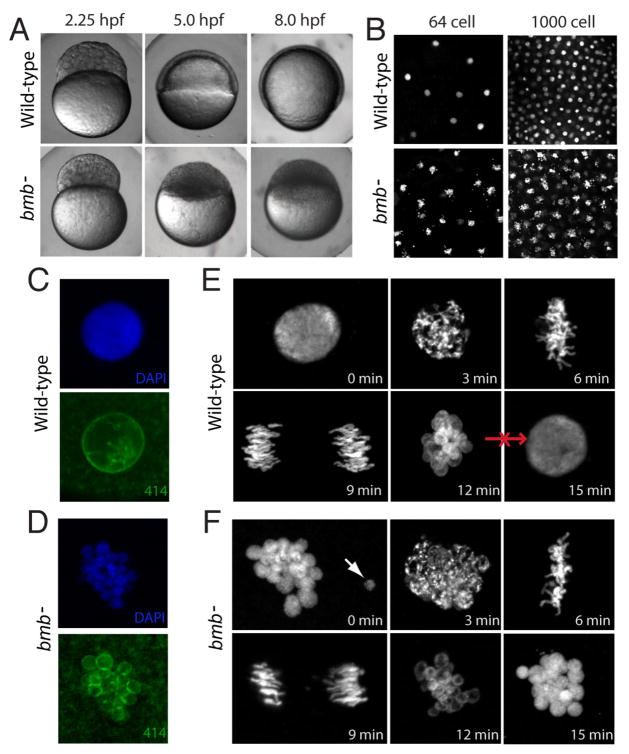
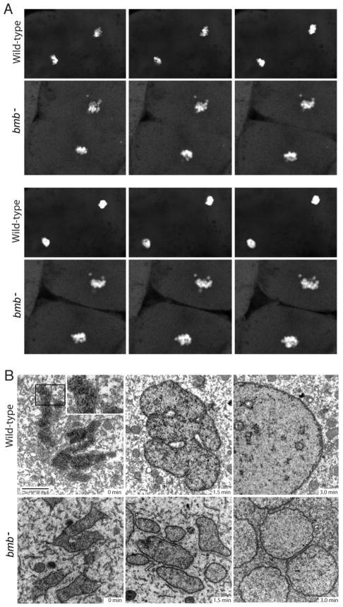
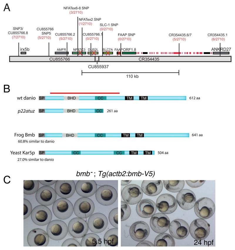
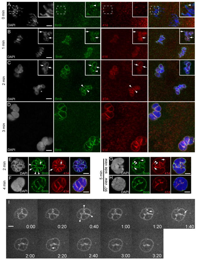
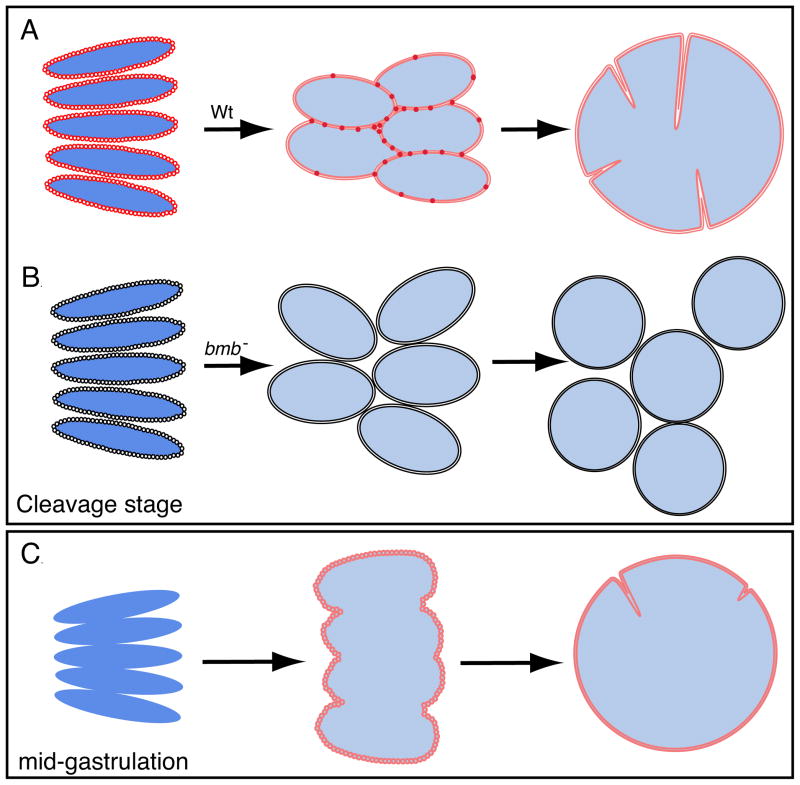
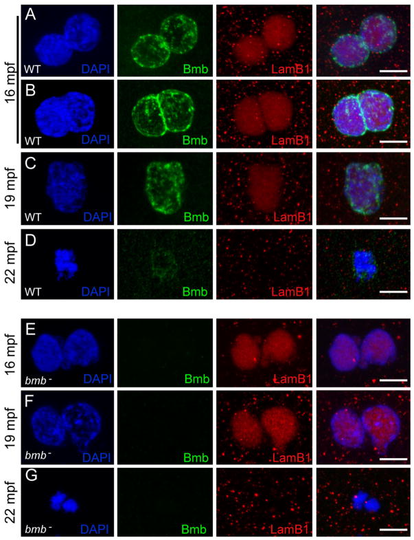
To accommodate the large cells following zygote formation, early blastomeres employ modified cell divisions. Karyomeres are one such modification, mitotic intermediates wherein individual chromatin masses are surrounded by nuclear envelope; the karyomeres then fuse to form a single mononucleus. We identified brambleberry, a maternal-effect zebrafish mutant that disrupts karyomere fusion, resulting in formation of multiple micronuclei. As karyomeres form, Brambleberry protein localizes to the nuclear envelope, with prominent puncta evident near karyomere-karyomere interfaces corresponding to membrane fusion sites. brambleberry corresponds to an unannotated gene with similarity to Kar5p, a protein that participates in nuclear fusion in yeast. We also demonstrate that Brambleberry is required for pronuclear fusion following fertilization in zebrafish. Our studies provide insight into the machinery required for karyomere fusion and suggest that specialized proteins are necessary for proper nuclear division in large dividing blastomeres.


Figures

Figure 1
Figure 1. bmb is required for early development and proper nuclear morphology
(A) Prior to the MBT (2.25 hpf), embryos from bmb mutant females are morphologically similar to WT embryos. After the MBT at 5.0 and 8.0 hpf, bmb mutants fail to undergo cell movements associated with epiboly and gastrulation. (B) DAPI staining of interphase nuclei during cleavage in WT (top) and bmb (bottom) reveals multiple chromatin bodies associated with each nucleus. (C) WT and (D) bmb interphase nuclei stained with mab414 (green) indicate that bmb nuclei are multi-micronucleated compared to WT. (E) WT and (F) bmb cell cycle time course at the 2 to 4 cell transition (images are projections of multiple confocal Z-slices). Top left –interphase; Top middle- prophase, Top right-metaphase, bottom left -anaphase, bottom middle- telophase, bottom right-interphase. In E and F for each time point, n=3. In this and subsequent figures ‘n’ refers to number of embryos examined (unless otherwise noted). In each case multiple nuclei or cells of each embryo were also examined.
Figure 2
Figure 2. Nuclear membrane fusion is disrupted in bmb mutants
(A) Frames from time-lapse experiments at the telophase-interphase transition demonstrate that chromatin bodies normally coalesce in WT (top) but fail to do so in bmb (bottom). Note: 6 frames were selected (from Movie S1 and S2 to best align the sequence of events between WT and bmb at the telophase to interphase transition. (B) Electron microscopy of WT versus bmb at the telophase-interphase transition. Embryos at the 128-cell stage were fixed at 90-second intervals for TEM. Black bar = 2 microns. The inset in WT (0 min) is enlarged 2X to show the double membrane nuclear envelope. For each time point n=2 embryos. Multiple cells from each embryo were examined in both WT and bmb.
Figure 3
Figure 3. bmb encodes a conserved novel predicted transmembrane protein
(A) Recombinants over total meiotic events are indicated parenthetically in red below the marker name. Candidate genes eliminated based on their WT cDNA sequence are indicated with a red X. A histone gene cluster is indicated as red boxes. Two predicted transposable elements are indicated by black boxes. (B) ORF1.8 encodes a novel 612 residue protein with a predicted N-terminal signal peptide sequence (SP) followed by a Bmb Homology Domain (BHD), a coiled-coil (CC) and two C-terminal transmembrane domains (TM) initiating at amino acid residues 363 and 415 (TMpred program) (Hofmann and Stoffel, 1993). Yeast Kar5p is 27% similar (excluding the C-terminal region) to the D. rerio homolog. The red bar corresponds to the recombinant protein used to generate Bmb antibodies. (C) Rescued bmb embryos at 50% epiboly (5.5 hpf) and 24 hpf. See also Figure S1.
Figure 4
Figure 4. Bmb protein dynamics during mitosis
(A) Bmb protein localizes as distinct foci to the mitotic spindle region during metaphase (n=4). (B) During anaphase Bmb is interspersed between the separating chromosomes (inset; n=3) and in the region of the mitotic spindle. (C) Later in anaphase, Bmb foci are decorating the chromosomes (inset; n=3). (D) At the 2 min time point Bmb surrounds the individual chromosomes, which are still separating (n=5). (E) Schematic summarizing Bmb localization during metaphase to anaphase/telophase. Arrows in the right panel indicate that the karyomeres will continue to move to their final central position in the cell. 0 min corresponds to metaphase at the 32-to-64 cell transition. Bmb (green), microtubules (DM1a, red), chromosomes (DAPI, white and blue in merge). Scale bar = 5 μm and insets show 2X enlargements. See also Figure S2.
Figure 5
Figure 5. Bmb foci mark sites corresponding to pre-, post- and active karyomere fusion
(A) At time point designated 0 min (mid-anaphase) at the 32- to 64-cell transition, Bmb protein (green) is first seen accumulating at the leading edge of the separating chromosomes (inset, arrowhead) prior to the assembly of nucleoporins (marked by mab414, red). (B) As the chromosomes continue to separate, nuclear membrane (marked by mab414) and Bmb surround individual chromosomes forming karyomeres. Insets in B show a more advanced example from the same time point (1 min). (C) Karyomeres become increasingly more spherical and prominent Bmb foci are more apparent at karyomere-karyomere interfaces (arrowheads). In another example, Bmb foci span a former interface, which now lacks nuclear membrane (inset, arrowheads) and a prominent Bmb aggregate interfaces adjacent karyomeres (inset, arrowhead). (D) At the 3 min time point larger secondary karyomeres are present. (E) Another 2 min example at a 90° view compared to C (an asterisk in C marks reference point). Bmb foci at karyomere-karyomere interfaces, where membrane is still present (arrowheads) or partially/completely absent (arrows). (F) Secondary karyomere formation at a 45° view. ( G) Bmb foci (Bmb, arrowheads) flank presumptive karyomere pre-fusion site (membrane still present- 414, arrowheads) and Bmb foci can be found at presumptive karyomere post-fusion sites (lack of membrane- 414, arrows). Note G–H shows mononucleus formation (5 min) from a lateral side view (G) and another sample from a 90° vantage point (reference point marked with asterisk in G) (H). All panels correspond to individual confocal Z-slices. Scale bar = 5 μm and insets show 2X enlargements. For each time point n ≥ 3. (I) Time-lapse imaging of Bmb-venus. Bmb foci are seen at the karyomere-karyomere interfaces (arrowheads) and are ultimately eliminated (arrows) as the mononucleus is formed. See also Movie S3 and Figure S3 for details.
Figure 6
Figure 6. Model of nuclear division during embryonic development
(A) During cleavage, the nuclear envelope and Bmb assemble on individual chromosomes as they continue to separate in mitosis resulting in karyomeres forming. As elongated karyomeres transition into more spherical shaped bodies, Bmb puncta becomes more prevalent at the karyomere-karyomere interfaces and fusion ensues to form a mononucleus. (B) In bmb mutants karyomeres still form, however, the absence of Bmb leads to a failure in membrane fusion resulting in multiple micronuclei forming. (C) At mid-gastrulation Bmb protein and the nuclear envelope do not assemble on individual chromosomes, instead, they assemble on a single chromatin mass and karyomere fusion is not necessary.
Figure 7
Figure 7. Bmb is required for pronuclear fusion
(A) At 16 mpf WT pronuclei are fully congressed. (B) Some WT pronuclei begin to fuse at 16 mpf. (C) At 19 mpf WT pronuclear fusion is complete. (D) At 22 mpf in WT embryos a single condensed DNA mass is detected at the first prometaphase. (E) At 16 mpf bmb pronuclei are fully congressed. (F) At 19 mpf bmb pronuclei fail to fuse as chromatin begins to condense. (G) At 22 min two separate condensed DNA masses are detected in bmb embryos at the first prometaphase. Scale bars = 10 μm. At each time point n ≥ 4. Images correspond to Z-projections of individual confocal Z-planes. WT TL males were crossed to female bmb mutants in E–G.

References

    1. Abramoff MD, Magalhaes PJ, JRS . Image Processing with ImageJ. Biophotonics International; 2004.
    1. Anderson DJ, Hetzer MW. Nuclear envelope formation by chromatin-mediated reorganization of the endoplasmic reticulum. Nat Cell Biol. 2007;9:1160–1166. - PubMed
    1. Anderson DJ, Hetzer MW. Reshaping of the endoplasmic reticulum limits the rate for nuclear envelope formation. J Cell Biol. 2008;182:911–924. - PMC - PubMed
    1. Balakier H, Cadesky K. The frequency and developmental capability of human embryos containing multinucleated blastomeres. Hum Reprod. 1997;12:800–804. - PubMed
    1. Baur T, Ramadan K, Schlundt A, Kartenbeck J, Meyer HH. NSF- and SNARE-mediated membrane fusion is required for nuclear envelope formation and completion of nuclear pore complex assembly in Xenopus laevis egg extracts. J Cell Sci. 2007;120:2895–2903. - PubMed

Publication types

MeSH terms

Associated data