. 2012 Aug 3;150(3):590-605.
doi: 10.1016/j.cell.2012.06.026.

Scl represses cardiomyogenesis in prospective hemogenic endothelium and endocardium

Affiliations

Scl represses cardiomyogenesis in prospective hemogenic endothelium and endocardium

Ben Van Handel et al. Cell. .

Abstract

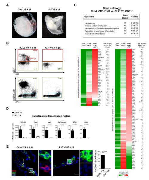
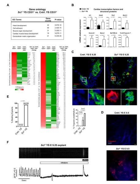
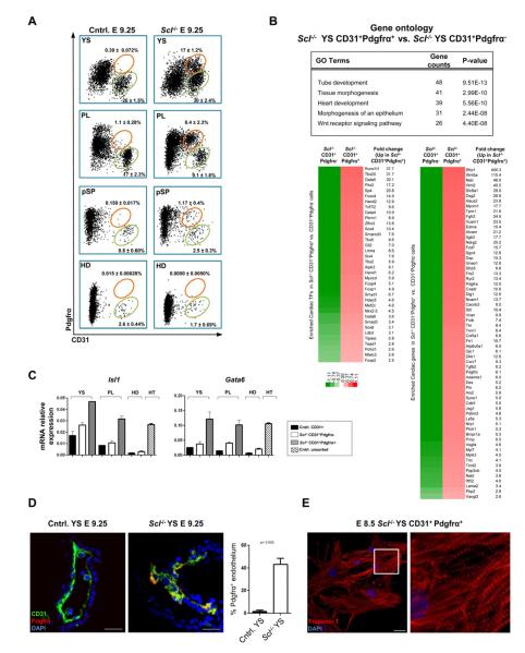
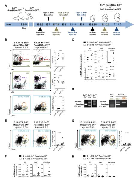
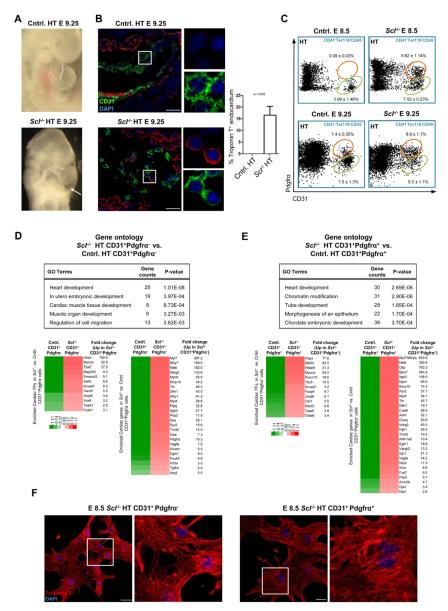
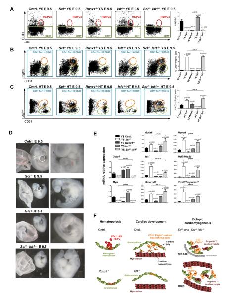
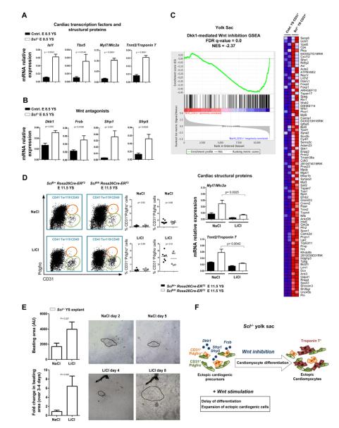
Endothelium in embryonic hematopoietic tissues generates hematopoietic stem/progenitor cells; however, it is unknown how its unique potential is specified. We show that transcription factor Scl/Tal1 is essential for both establishing the hematopoietic transcriptional program in hemogenic endothelium and preventing its misspecification to a cardiomyogenic fate. Scl(-/-) embryos activated a cardiac transcriptional program in yolk sac endothelium, leading to the emergence of CD31+Pdgfrα+ cardiogenic precursors that generated spontaneously beating cardiomyocytes. Ectopic cardiogenesis was also observed in Scl(-/-) hearts, where the disorganized endocardium precociously differentiated into cardiomyocytes. Induction of mosaic deletion of Scl in Scl(fl/fl)Rosa26Cre-ER(T2) embryos revealed a cell-intrinsic, temporal requirement for Scl to prevent cardiomyogenesis from endothelium. Scl(-/-) endothelium also upregulated the expression of Wnt antagonists, which promoted rapid cardiomyocyte differentiation of ectopic cardiogenic cells. These results reveal unexpected plasticity in embryonic endothelium such that loss of a single master regulator can induce ectopic cardiomyogenesis from endothelial cells.


Figures

Figure 1
Figure 1. Scl establishes hemogenic endothelium in the yolk sac
(A) Analysis of E 9.25 Scl−/− embryos documented lack of primitive red cells (arrow) and (B) CD41+cKit+ HS/PCs. (B) Sorting strategy used to isolate endothelial cells from control and Scl−/− yolk sacs (YS). Cells lacking hematopoietic markers CD41 (green boxes), CD45 and Ter119 but expressing CD31+ (blue boxes) were subjected to gene expression analysis. (C) Heat maps represent the relative expression and fold change of hematopoietic transcription factors (TFs) and other proteins significantly downregulated (>1.5 fold, p<0.05) in Scl−/− vs. control yolk sac endothelium; expression in control CD41+cKit+ HS/PCs is shown for comparison. Selected GO (gene ontology) categories derived from differentially expressed genes are listed. (D) qPCR for hematopoietic TFs on E 9.25 unfractionated yolk sacs. Mean ± SEM of 4 biological replicates normalized to HPRT is shown. (E) IF for Scl dependent HS/PC marker CD41 (n=3); insets are higher magnification, single channel + DAPI images of boxed areas. Scale bar represents 25 μm.
Figure 2
Figure 2. Lack of Scl induces cardiomyogenesis in yolk sac vasculature
(A) Selected GO (gene ontology) categories encompassing genes upregulated in Scl−/− yolk sac endothelium are shown. Heat maps represent the relative expression and fold change of known cardiac transcription factors (TFs) and other proteins significantly upregulated (>1.5 fold, p<0.05) in Scl−/− vs. control yolk sac CD31+ cells. CD41+cKit+ HS/PCs are shown for comparison. (B) qPCR for cardiac TFs and cardiomyocyte structural genes on E 9.25 unfractionated yolk sacs. Mean ± SEM of 4 biological replicates normalized to HPRT is shown. (C) IF for Troponin I (n=2; Troponin T n=3) and CD31 on yolk sac sections; insets are higher magnification, single channel + DAPI images of boxed areas. Scale bar denotes 25 μm. (D) Whole mount IF for Troponin T on yolk sacs. Scale bar denotes 50 μm. (E) Frequency of yolk sacs generating beating colonies in explant culture (see Movie S1). (F) Scl−/− yolk sac explants were loaded with Fluo4-AM and spontaneous calcium transients were measured using confocal microscopy. Nifedipine (10μM) and caffeine (10mM) were added at the indicated times.
Figure 3
Figure 3. Scl represses the development of CD31+Pdgfrα+ cardiac cells in hemogenic tissues
(A) FACS on cells depleted for CD41, CD45 and Ter119 showing CD31+Pdgfrα+ cells (orange gates) and CD31+ endothelial cells (green gates) in embryonic tissues (YS, yolk sac; PL, placenta; pSP, para-aortic splanchnopleura, HD, head). Mean ± SEM of at least 3 biological replicates is shown. (B) Gene expression analysis of CD31+Pdgfrα−/+ cells from Scl−/− yolk sacs. Differentially expressed (>1.5 fold, p<0.05) cardiac transcription factors (TFs) and other proteins enriched in CD31+Pdgfrα+ cells with their fold changes, as well as selected GO (gene ontology) categories, are shown. (C) qPCR for cardiac TFs on indicated populations sorted from YS, PL and HD; unfractionated heart (HT) was used as a positive control. Mean ± SD of technical replicates from pooled samples normalized to actin is shown. (D) IF for CD31 and Pdgfrα on yolk sac sections (n=3). Scale bar represents 25 μm.(See Movie S2). (E) CD31+Pdgfrα+ cells were sorted from E 8.5 Scl−/− yolk sacs, cultured and stained for Troponin T. Scale bar denotes 25 μm.
Figure 4
Figure 4. Scl has a cell autonomous, temporally defined role in preventing ectopic cardiogenesis in hemogenic tissues
(A) Tamoxifen-inducible Rosa26Cre-ERT2 mouse strain was used to delete Scl at desired time points. (B, E, G) FACS for CD41+cKit+ HS/PCs (red ovals) and Ter119+ RBCs (red blood cells; purple circles) in Rosa26Cre-ERT2 Sclfl/+ and Rosa26Cre-ERT2 Sclfl/fl yolk sacs of pregnant dams injected at E 6.5 (B), E 7.5 (E) and F 8.5 (G). Analysis for CD31+ cells (green ovals) and CD31+Pdgfrα+ cells (orange circles) is shown. Graphs show frequencies of HS/PCs, RBCs and CD31+Pdgfrα+ cells in individual embryos. (C, F, H) qPCR for cardiac and/or hematopoietic genes on yolk sacs following injection at E 6.5 (C), E 7.5 (F) and (H) E 8.5. Graphs show mRNA expression level normalized to HPRT in individual embryos. (D) PCR of embryonic head DNA (left) isolated from embryos injected at E 6.5 and harvested at E 9.5. PCR from CD31+Pdgfrα vs. CD31+Pdgfrα+ cells sorted from Rosa26Cre-ERT2 Sclfl/fl yolk sacs (right).
Figure 5
Figure 5. Scl−/− endocardium is misspecified to cardiomyogenic fate
(A) Scl−/− hearts evidenced malformed outflow tract (dotted lines) and pericardial effusion (arrow). (B) IF for CD31 and Troponin T on heart sections (n=3). Scale bar represents 25 μm. (C) FACS for CD31+ endocardial cells (green ovals) and CD31+Pdgfrα+ cells (orange circles) in the heart at E 8.5 and E 9.25. Mean ± SEM of at least 3 biological replicates is shown. (D) Gene expression analysis of Scl−/− and control CD31+Pdgfrα endocardial cells. Differentially expressed (>1.5 fold, p<0.05) cardiac transcription factors (TFs) and other proteins and their fold changes, as well as selected GO (gene ontology) categories enriched in Scl−/− CD31+Pdgfrα cells, are shown. (E) Gene expression analysis of Scl−/− and control CD31+Pdgfrα+ cells from the heart. Differentially expressed (>1.5 fold, p<0.05) cardiac TFs and other proteins and their fold changes, as well as selected GO categories enriched in Scl−/− CD31+Pdgfrα+ cells, are shown. (F) IF for Troponin T on cells derived from E 8.5 Scl−/− CD31+Pdgfrα−/+ cells that were sorted and cultured (see Movie S3). Scale bar denotes 25 μm.
Figure 6
Figure 6. Runx1 and Isl1 are not major regulators of the ectopic cardiomyogenic cells
FACS was performed on yolk sac (A and B) and hearts (C) isolated from control, Scl−/−, Runx1−/−, Isl1−/− and Scl−/− Isl1−/− embryos. All p values shown are with respect to control unless otherwise indicated. (A) CD41+cKit+ HS/PCs (red ovals) are shown in yolk sac and CD31+Pdgfrα+ cells (orange ovals) are shown in yolk sac (B) and heart (C). (D) Scl−/− Isl1−/− embryos lacked blood in the yolk sac (arrows, left panels), pharyngeal arches (arrowheads, left panels) and outflow tract (circles, left panels). (E) qPCR for hematopoietic and cardiac transcription factors and cardiomyocyte structural genes on E 9.5 unfractionated yolk sacs. Mean ± SEM of at least 2 (Runx1−/− = 2; all others ≥5) biological replicates normalized to HPRT is shown. (F) Runx1 and Isl1 are required for HS/PC generation and cardiac cushion development respectively; however, neither regulates ectopic cardiogenesis.
Figure 7
Figure 7. Scl−/− endothelium expresses and responds to Wnt antagonists that promote cardiomyocyte differentiation
(A) qPCR for cardiac transcription factors and cardiomyocyte structural genes and (B) Wnt antagonists on E 8.5 unfractionated yolk sacs. Mean ± SEM of 4 biological replicates normalized to HPRT is shown. (C) GSEA of expression data from E 9.25 Scl−/− and control yolk sac CD31+ cells compared to embryoid bodies treated with the Wnt antagonist Dkk1. The heat map depicts genes that are part of the core enrichment set. The False Discovery Rate (FDR) q-value and Normalized Enrichment Score (NES) are shown. (D) Pregnant dams were injected with tamoxifen on E 6.5 and treated daily with either NaCl (control) or LiCl (Wnt agonist). FACS analysis showing the frequency of CD31+Pdgfrα and CD31+Pdgfrα+ cells after NaCl or LiCl treatment and qPCR for Myl7 and Tnnt2. (E) Scl−/− yolk sacs were cultured with either NaCl or LiCl; videos of beating clusters were taken and the area measured in arbitrary units (AU; n ≥27). Explants were imaged at two time points during the culture period (n ≥3); beating areas are outlined. (F) Schematic depicting the acceleration of differentiation of ectopic cardiogenic cells by Wnt inhibitors derived from Scl−/− endothelium; this could be attenuated by the Wnt agonist LiCl.

References

    1. Ahnfelt-Ronne J, Jorgensen MC, Hald J, Madsen OD, Serup P, Hecksher-Sorensen J. An improved method for three-dimensional reconstruction of protein expression patterns in intact mouse and chicken embryos and organs. The journal of histochemistry and cytochemistry : official journal of the Histochemistry Society. 2007;55:925–930. - PubMed
    1. Alva JA, Zovein AC, Monvoisin A, Murphy T, Salazar A, Harvey NL, Carmeliet P, Iruela-Arispe ML. VE-Cadherin-Cre-recombinase transgenic mouse: a tool for lineage analysis and gene deletion in endothelial cells. Developmental dynamics : an official publication of the American Association of Anatomists. 2006;235:759–767. - PubMed
    1. Bertrand JY, Chi NC, Santoso B, Teng S, Stainier DY, Traver D. Haematopoietic stem cells derive directly from aortic endothelium during development. Nature. 2010;464:108–111. - PMC - PubMed
    1. Bertrand JY, Giroux S, Golub R, Klaine M, Jalil A, Boucontet L, Godin I, Cumano A. Characterization of purified intraembryonic hematopoietic stem cells as a tool to define their site of origin. Proc Natl Acad Sci U S A. 2005;102:134–139. - PMC - PubMed
    1. Boisset JC, van Cappellen W, Andrieu-Soler C, Galjart N, Dzierzak E, Robin C. In vivo imaging of haematopoietic cells emerging from the mouse aortic endothelium. Nature. 2010;464:116–120. - PubMed

Publication types

MeSH terms

Substances

Associated data