. 2012 Nov;103(11):1946-54.
doi: 10.1111/j.1349-7006.2012.02408.x. Epub 2012 Sep 14.

Hypoxia induces gefitinib resistance in non-small-cell lung cancer with both mutant and wild-type epidermal growth factor receptors

Affiliations

Hypoxia induces gefitinib resistance in non-small-cell lung cancer with both mutant and wild-type epidermal growth factor receptors

Kunihiko Minakata et al. Cancer Sci. 2012 Nov.

Abstract

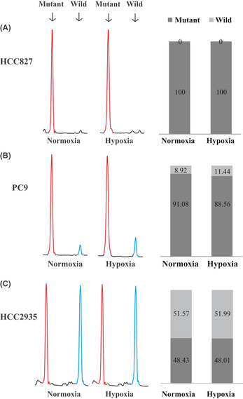
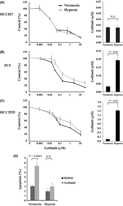
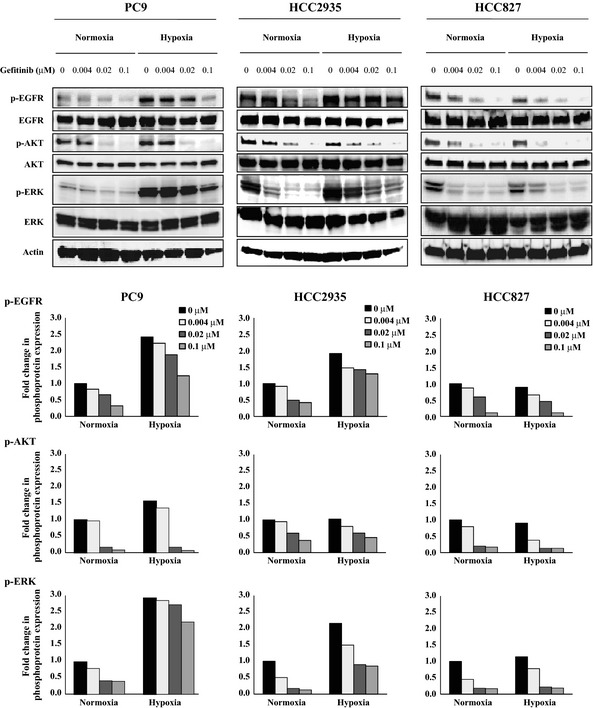
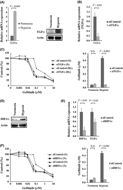
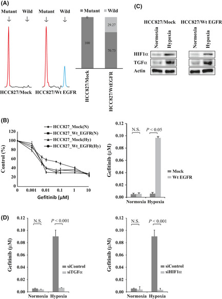
Somatic mutations in the epidermal growth factor receptor (EGFR) gene, such as exon 19 deletion mutations, are important factors in determining therapeutic responses to gefitinib in non-small-cell lung cancer (NSCLC). However, some patients have activating mutations in EGFR and show poor responses to gefitinib. In this study, we examined three NSCLC cell lines, HCC827, PC9, and HCC2935, that expressed an EGFR exon 19 deletion mutation. All cells expressed mutant EGFR, but the PC9 and HCC2935 cells also expressed wild-type EGFR. The HCC827 cells were highly sensitive to gefitinib under both normoxia and hypoxia. However, the PC9 and HCC2935 cells were more resistant to gefitinib under hypoxic conditions compared to normoxia. Phosphorylation of EGFR and ERK was suppressed with gefitinib treatment to a lesser extent under hypoxia. The expression of transforming growth factor-α (TGFα) was dramatically upregulated under hypoxia, and the knockdown of TGFα or hypoxia-inducible factor-1α (HIF1α) reversed the resistance to gefitinib in hypoxic PC9 and HCC2935 cells. Finally, introduction of the wild-type EGFR gene into the HCC827 cells caused resistance to gefitinib under hypoxia. This phenomenon was also reversed by the knockdown of TGFα or HIF1α. Our results indicate that hypoxia causes gefitinib resistance in EGFR-mutant NSCLC through the activation of wild-type EGFR mediated by the upregulation of TGFα. The presence of wild-type and mutant EGFR along with tumor hypoxia are important factors that should be considered when treating NSCLC patients with gefitinib.


Figures

Figure 1
Figure 1
Genetic status of epidermal growth factor receptor (EGFR) in HCC827 (A), PC9 (B), and HCC2935 (C) non‐small‐cell lung carcinoma cell lines. Left: Electropherograms of fragment analysis under normoxia and hypoxia. Red peak, amplified PCR fragment of exon 19 deletion mutation EGFR (73 or 70 bp); blue peak, wild‐type EGFR (88 bp). Right: Relative proportions of mutant and wild‐type EGFR expression determined by fragment analysis. Black bar, mutant EGFR; gray bar, wild‐type EGFR.
Figure 2
Figure 2
(A–C) Hypoxia induced gefitinib resistance in PC9 (B) and HCC2935 (C) but not in HCC827 (A) non‐small‐cell lung carcinoma cells. Left: Cells were incubated under hypoxia (1% O 2) or normoxia (21% O 2) for 48 h, followed by treatment with the indicated concentrations of gefitinib for 48 h. Each data point represents the average value of four samples and is expressed as a percentage of the surviving cells relative to the untreated controls. Right: Bar graphs showing the IC 50 values of cells resistant to gefitinib. (D) After hypoxia or normoxia for 48 h, PC9 cells were grown in the presence of 0.1% DMSO (control) or 0.1 μM gefitinib for 24 h. Apoptosis was assessed using propidium iodide and annexin‐V staining. The y‐axis denotes the sum of the early and late apoptotic cells as the mean ± standard error of the mean (n = 3). N.S., not significant.
Figure 3
Figure 3
Effects of gefitinib treatment on the levels of phosphorylated epidermal growth factor receptor (EGFR), AKT, and ERK (p‐EGFR, p‐AKT, and p‐ERK, respectively) under normoxia and hypoxia. The PC9, HCC2935, and HCC827 non‐small‐cell lung carcinoma cells were incubated under hypoxia or normoxia for 48 h, then either left untreated or treated with the indicated concentrations of gefitinib for 3 h. After treatment, the cells were lysed and equal amounts of cell lysates were subjected to Western blot analysis using antibodies against total and phosphorylated EGFR (Y‐1068), AKT, and ERK. The levels of actin served as internal controls for equal protein loading in each lane. The bar graph below each blot shows the fold change in phospho‐protein expression due to treatment with the indicated concentrations of gefitinib. The fold changes were calculated by setting the ratios of the phospho‐protein/total protein band intensities for the untreated normoxia cells to unity.
Figure 4
Figure 4
Transforming growth factor‐α (TGFα) and hypoxia‐inducible factor‐1α (HIF1α) were upregulated by hypoxia, and TGFα or HIF1α knockdown reversed hypoxia‐induced resistance to gefitinib in PC9 non‐small‐cell lung carcinoma cells. (A) TGFα messenger RNA and protein expression in PC9 cells under normoxia and hypoxia were analyzed by quantitative RTPCR (left) and Western blotting (right). (B) TGFα expression was knocked down in PC9 cells with siRNA. Two specific siRNAs and one non‐specific control were used, and data representative of the siRNA experiment are shown (P = 0.008). (C) Left: PC9 cells were transfected with siTGFα or siControl then incubated under hypoxia (Hy) or normoxia (N) for 48 h, followed by treatment with the indicated concentrations of gefitinib. Each data point is expressed as a percentage of the surviving cells relative to the untreated controls. Right: Bar graphs show the IC 50 values of the cells to gefitinib. The knockdown of TGFα expression significantly reduced the IC 50 value of the hypoxic PC9 cells. P < 0.001. (D) HIF1α expression in PC9 cells under normoxia and hypoxia was analyzed by Western blotting. (E) HIF1α expression was knocked down with siHIF1α in PC9 cells, resulting in the downregulation of TGFα expression (P = 0.004, P = 0.03, respectively). Two specific siRNAs and one non‐specific control were used; data representative of the siRNA experiment is shown. (F) Left: PC9 cells were transfected with siHIF1α or siControl then incubated under hypoxia (Hy) or normoxia (N) for 48 h followed by treatment with the indicated concentrations of gefitinib. Right: Bar graphs show the IC 50 values of cells to gefitinib. The knockdown of HIF1α expression significantly reduced the IC 50 value of the hypoxic PC9 cells. P < 0.001.
Figure 5
Figure 5
Transfection of wild‐type epidermal growth factor receptor (EGFR) gene into HCC827 cells harboring the activating EGFR mutation. (A) Transfectants of the empty vector and wild‐type EGFR were designated HCC827/Mock and HCC827/Wt EGFR cells, respectively. Left: Electropherograms of the fragment analysis for the HCC827/Mock and HCC827/Wt EGFR cells. Red peak, amplified PCR fragment of exon 19 deletion mutation EGFR; blue peak, wild‐type EGFR. Right: Relative proportions of mutant and wild‐type EGFR expression by fragment analysis. Black bar, mutant EGFR; gray bar, wild‐type EGFR. (B) HCC827/Mock and HCC827/Wt EGFR cells were incubated under hypoxia or normoxia for 48 h followed by treatment with gefitinib. The IC 50 value of the HCC827/Wt EGFR cells was significantly higher than that of the HCC827/Mock cells under hypoxia. P = 0.014. (C) HIF1α and TGFα expression was upregulated by hypoxia in the HCC827/Mock cells (left) and the HCC827/Wt EGFR cells (right) as indicated by Western blotting. (D) HCC827/Wt EGFR cells were transfected with siTGFα or siControl (left) or with siHIF1α or siControl (right) then incubated under hypoxia or normoxia for 48 h, followed by treatment with gefitinib. The IC 50 values of the HCC827/Wt EGFR cells were significantly reduced by the knockdown of TGFα or HIF1α under hypoxia (P < 0.001, P < 0.001, respectively).

References

    1. Jemal A, Siegel R, Xu J, Ward E. Cancer statistics, 2010. CA Cancer J Clin 2010; 60: 277–300. - PubMed
    1. Ohsaki Y, Tanno S, Fujita Y et al Epidermal growth factor receptor expression correlates with poor prognosis in non‐small cell lung cancer patients with p53 overexpression. Oncol Rep 2000; 7: 603–7. - PubMed
    1. Hirsch FR, Varella‐Garcia M, Bunn PA Jr et al Epidermal growth factor receptor in non‐small‐cell lung carcinomas: correlation between gene copy number and protein expression and impact on prognosis. J Clin Oncol 2003; 21: 3798–807. - PubMed
    1. Sharma SV, Bell DW, Settleman J, Haber DA. Epidermal growth factor receptor mutations in lung cancer. Nat Rev Cancer 2007; 7: 169–81. - PubMed
    1. Rusch V, Baselga J, Cordon‐Cardo C et al Differential expression of the epidermal growth factor receptor and its ligands in primary non‐small cell lung cancers and adjacent benign lung. Cancer Res 1993; 53: 2379–85. - PubMed

Publication types

MeSH terms