Comparative Study
. 2012 Aug 3:12:166.
doi: 10.1186/1471-2180-12-166.

Pathogenic Mycobacterium bovis strains differ in their ability to modulate the proinflammatory activation phenotype of macrophages

Affiliations
Comparative Study

Pathogenic Mycobacterium bovis strains differ in their ability to modulate the proinflammatory activation phenotype of macrophages

Marcelle Rm Andrade et al. BMC Microbiol. .

Abstract

Background: Tuberculosis, caused by Mycobacterium tuberculosis or Mycobacterium bovis, remains one of the leading infectious diseases worldwide. The ability of mycobacteria to rapidly grow in host macrophages is a factor contributing to enhanced virulence of the bacteria and disease progression. Bactericidal functions of phagocytes are strictly dependent on activation status of these cells, regulated by the infecting agent and cytokines. Pathogenic mycobacteria can survive the hostile environment of the phagosome through interference with activation of bactericidal responses. To study the mechanisms employed by highly virulent mycobacteria to promote their intracellular survival, we investigated modulating effects of two pathogenic M. bovis isolates and a reference M. tuberculosis H37Rv strain, differing in their ability to multiply in macrophages, on activation phenotypes of the cells primed with major cytokines regulating proinflammatory macrophage activity.

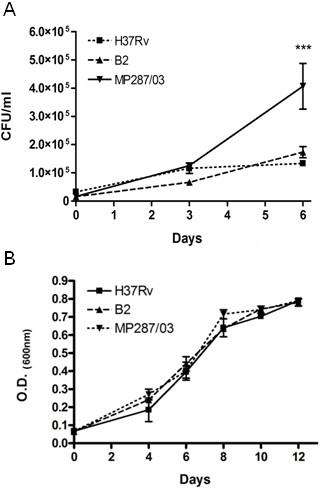
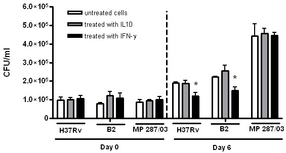
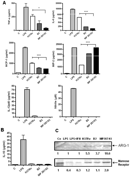
Results: Bone marrow- derived macrophages obtained from C57BL/6 mice were infected by mycobacteria after a period of cell incubation with or without treatment with IFN-γ, inducing proinflammatory type-1 macrophages (M1), or IL-10, inducing anti-inflammatory type-2 cells (M2). Phenotypic profiling of M1 and M2 was then evaluated. The M. bovis strain MP287/03 was able to grow more efficiently in the untreated macrophages, compared with the strains B2 or H37Rv. This strain induced weaker secretion of proinflammatory cytokines, coinciding with higher expression of M2 cell markers, mannose receptor (MR) and arginase-1 (Arg-1). Treatment of macrophages with IFN-γ and infection by the strains B2 and H37Rv synergistically induced M1 polarization, leading to high levels of inducible nitric oxide synthase (iNOS) expression, and reduced expression of the Arg-1. In contrast, the cells infected with the strain MP287/03 expressed high levels of Arg-1 which competed with iNOS for the common substrate arginine, leading to lower levels of NO production.

Conclusions: The data obtained demonstrated that the strain, characterized by increased growth in macrophages, down- modulated classical macrophage activation, through induction of an atypical mixed M1/M2 phenotype.


Figures

Figure 1
Figure 1
Evaluation of the growth properties of M. bovis isolates. Isolates obtained from animals with tuberculosis, strains MP287/03 and B2, and reference M. tuberculosis strain H37Rv, were used for infection of BMDM in vitro (A) or cultured in Middlebrook 7H9 broth (B). Growth rates of mycobacteria inside MΦ infected at MOI of 1 were determined using the colony count method. Intracellular CFU numbers were quantified immediately after infection (day 0) or at 3 or 6 days after infection (A). Growth rates of mycobacteria in 7 H9 Middlebrook broth were monitored by measurement of OD of the mycobacterial cultures by spectrophotometry. The growth curves of the mycobacterial strains within a 12 day period of incubation are presented. (B). Values are the means ± SD of three independent experiments with samples in triplicate.
Figure 2
Figure 2
The capacity of pathogenic mycobacteria to grow intracellularly in macrophages treated with IFN-γor IL-10. Cultures of BMDM were pretreated with exogenic murine r-IFN -γ or r-IL-10 for 2 h, infected with the mycobacterial strains at a MOI of 1, as indicated in the legend to Figure 1, and incubated in the presence of these cytokines for an additional 6 days. The intracellular CFU numbers determined at day 0 and day 6 are presented. The data of three independent experiments are shown as mean ± SD of samples in triplicate. Asterisks represent statistical significance (p < 0.05) compared to infected cells cultured without addition of the cytokines.
Figure 3
Figure 3
The activation profiles of macrophages infected with pathogenic mycobacteria. BMDM were infected with the studied mycobacterial strains at a MOI of 5:1, washed and incubated for an additional 48 h. The cells left untreated and cells stimulated with LPS for 48 h were used as a negative and positive controls of proinflammatory activation, respectively. To evaluate markers of M1-type activation (A), the culture supernatants of infected cultures were harvested and tested for TNF-α, IL-6, MCP-1, MIP-2 and IL-12 by Bioplex test, and for NO production by Griess reaction. Assays were completed with duplicate samples, and results are expressed as a mean of three independent experiments. Lines over bars indicate the isolates for which the induced cytokine production differed significantly from that induced by H37Rv (*p < 0.05; ***p < 0.001). To evaluate markers of M2-type activation, secretion of IL-10 was quantified by Bioplex assay (B), and expression of Arginase 1 and MR/CD206 in the adhered cells was tested by Western blotting (C). Lower panel, quantification of the protein levels by densitometric analysis of immunoreactive bands.
Figure 4
Figure 4
The activation profiles of macrophages treated with IFN-γ or IL-10 and infected with pathogenic mycobacteria. BMDM were pretreated, or not, with murine r-IFN-γ or r-IL-10 for 2 h, infected with the studied mycobacterial strains at a MOI of 5:1, washed, treated again with the cytokines and incubated for an additional 48 h. The cells stimulated with LPS and r-IFN-γ for 48 h, or left untreated, were used as a positive and negative controls of classical proinflammatory activation, respectively. To evaluate markers of M1-type activation, the culture supernatants were tested for proinflammatory mediator levels (A-C) and the adhered cells were tested for expression of iNOS (D). Measurement of TNF-α, IL-6, MCP-1, MIP-2 and IL-12 concentrations was performed by Bioplex test, and NO production was evaluated by Griess reaction Assays were completed with duplicate samples, and results are expressed as a mean of three independent experiments. To evaluate markers of M2-type activation, expression of Arginase 1 and MR/CD206 in the adhered cells was tested by Western blotting (E) and secretion of IL-10 was quantified by Bioplex assay (F). Lower panels in D and E, quantification of the protein levels by densitometric analysis of immunoreactive bands. Asterisks in A, B and F indicate the infected cultures treated with recombinant IFN-γ or IL-10, for which the induced cytokine production differed significantly from that in the corresponding cultures incubated without the presence of exogenic cytokines (*p < 0.05; **p < 0.01; ***p < 0.001). Lines over bars in A and B indicate the Mbv isolates for which the induced cytokine or NO production differed significantly from that induced by H37Rv (#p < 0.01; ##p < 0.001).

References

    1. Gordon S. Alternative activation of macrophages. Nat Rev Immunol. 2003;3:23–35. doi: 10.1038/nri978. - DOI - PubMed
    1. Mantovani A, Sica A, Sozzani S, Allavena P, Vecchi A, Locati M. The chemokine system in diverse forms of macrophage activation and polarization. Trends Immunol. 2004;25:677–686. doi: 10.1016/j.it.2004.09.015. - DOI - PubMed
    1. Mosser DM, Edwards JP. Exploring the full spectrum of macrophage activation. Nat Rev Immunol. 2008;8:958–969. doi: 10.1038/nri2448. - DOI - PMC - PubMed
    1. Lopez-Castejón G, Baroja-Mazo A, Pelegrín P. Novel macrophage polarization model: from gene expression to identification of new anti-inflammatory molecules. Cell Mol Life Sci. 2011;68:3095–3107. doi: 10.1007/s00018-010-0609-y. - DOI - PMC - PubMed
    1. Kleinnijenhuis J, Oosting M, Joosten LA, Netea MG, Van Crevel R. Innate immune recognition of Mycobacterium tuberculosis. Clin Dev Immunol. 2011;201(1):405310. - PMC - PubMed

Publication types