Comparative Study
. 2012 Oct;279(19):3715-3726.
doi: 10.1111/j.1742-4658.2012.08733.x. Epub 2012 Aug 31.

Homeodomain-containing protein HOXB9 regulates expression of growth and angiogenic factors, facilitates tumor growth in vitro and is overexpressed in breast cancer tissue

Affiliations
Comparative Study

Homeodomain-containing protein HOXB9 regulates expression of growth and angiogenic factors, facilitates tumor growth in vitro and is overexpressed in breast cancer tissue

Bishakha Shrestha et al. FEBS J. 2012 Oct.

Abstract

HOXB9 is a homeobox-containing gene and is critical for the development of mammary gland and sternum. HOXB9 is also regulated by estrogen and is critical for angiogenesis. We investigated the biochemical roles of HOXB9 and its homeodomain in cell-cycle progression and tumorigenesis. Our studies demonstrated that HOXB9 is overexpressed in breast cancer tissue. HOXB9 overexpression stimulated 3D formation in soft agar assay. HOXB9 binds to the promoters of various tumor growth and angiogenic factors and regulates their expression. The homeodomain of HOXB9 plays crucial roles in transcriptional regulation of tumor growth factors and also in 3D colony formation, indicating crucial roles of the HOXB9 homeodomain in tumorigenesis. Overall, we demonstrated that HOXB9 is a critical regulator of tumor growth factors and is associated with tumorigenesis.


Figures

Figure 1
Figure 1
HOXB9 expression in different human cell lines and breast cancer tissue. (A) The total RNA was isolated from different cell lines, reverse-transcribed to cDNA and analyzed by real-time PCR using primers specific to HOXB9. GAPDH was used as a loading control. HOXB9 expression relative to GAPDH is plotted. Each experiment was repeated at least thrice. Bars indicate standard errors (n=3, p<0.05)). (B-D) HOXB9 expression in breast cancer tissue: Human breast cancer tissue microarray (6 cases of breast cancer along with their matched adjacent normal breast tissue) was immunostained (DAB staining) with HOXB9 antibody. Magnified view of tissue histology (at two different magnification) showing HOXB9 expression in case 6 is shown in panel C and the relative quantification of HOXB9 expression within the tissue section is presented in panel D. Statistical analysis of 6 control and 6 breast cancer tissues showing the cumulative expression level of the control and cancer tissues is also shown (n= 6, p<0.05)
Figure 2
Figure 2
Subcellular distribution of HOXB9 and homeodomain truncated HOXB9. (A) Diagrammatic representation of Flag-HOXB9 and Flag-HOXB9-ΔHD. (B) Stable cells expressing Flag-HOXB9 and Flag-HOXB9-ΔHD were fractionated to cytoplasmic and nuclear extracts and analyzed by Western blotting using anti-flag antibody. (C) Flag-HOXB9 and Flag-HOXB9-ΔHD stable cells were immunostained with Flag and RNAPII antibodies followed by FITC or TRITC conjugated secondary antibodies. DAPI was used to stain the nucleus and visualized under a fluorescence microscope.
Figure 3
Figure 3
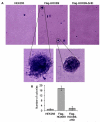
Colony forming ability of Flag-HOXB9 and Flag-HOXB9-ΔHD cells. HEK293 cells, Flag-HOXB9 and Flag-HOXB9-ΔHD overexpressed cells (stable cell lines) were grown in soft agar for 21 days and fed with media every 2 days. The colonies formed in soft agar were stained with 0.005% crystal violet solution and observed under microscope (panel A). Magnified views of selected colonies are shown on the bottom panel (A). Number of colonies grown in soft agar were counted under light microscope and plotted. Bars represent standard errors (n = 3, p<0.05).
Figure 4
Figure 4
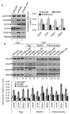
Role of HOXB9 in expression of tumor growth and angiogenic factors (A) Analysis of expression of growth and angiogenic factors. The RNA extracts of HEK293, Flag-HOXB9 and Flag-HOXB9-ΔHD over expressed cells were subjected to RT-PCR with primers specific to HOXB9, VEGF, bFGF, TGFβ1 and NRG2 and GAPDH (loading control). Real-time quantification is shown in right panel. Bars indicate statdard error ((n = 3, p<0.05). (B) ChIP assay: HEK293, Flag-HOXB9 and Flag-HOXB9-ΔHD cells were subjected to ChIP assay with anti-Flag, RNAPII and H3K4-trimethyl antibodies and the ChIP DNA was anlayzed by PCR with primers specific to promoters of VEGF, bFGF, TGFβ1 and NRG2. Real-time quantification of recruitment level of Flag-HOXB9 (relative to input) is on bottom panel. Bars indicate standard errors ((n = 3, p<0.05).
Figure 5
Figure 5
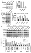
A-B) Effect of HOXB9 knockdown on the expression of tumor growth and angiogenic factors. HEK293 cells were transfected with HOXB9 specific and scramble antisenses (72 h). The protein extracts of control and antisense-treated cells were analyzed by Western blotting with HOXB9 and actin (loading control) antibodies (panel A, quantification on the bottom panel). The RNA from the control and antisense-treated cells were analyzed by RT-PCR with primers specific to HOXB9, VEGF, bFGF, TGFβ1 and NRG2 (panel B). GAPDH was used as control. Real time quantification of expression relative to GAPDH is shown in right panel. Bars indicate standard errors (n = 3, p<0.05). (C) ChIP assay: The control, scramble and HOXB9 antisense treated cells were subjected to ChIP assay with RNAPII, HOXB9, and H3K4-trimethyl antibodies. The ChIP DNA was PCR-amplified using primers specific to VEGF, bFGF, TGFβ1 and NRG2.

References

    1. Zakany J, Duboule D. The role of Hox genes during vertebrate limb development. Curr Opin Genet Dev. 2007;17:359–66. - PubMed
    1. Lappin TR, Grier DG, Thompson A, Halliday HL. HOX genes: seductive science, mysterious mechanisms. Ulster Med J. 2006;75:23–31. - PMC - PubMed
    1. Grier DG, Thompson A, Kwasniewska A, McGonigle GJ, Halliday HL, Lappin TR. The pathophysiology of HOX genes and their role in cancer. J Pathol. 2005;205:154–71. - PubMed
    1. Alexander T, Nolte C, Krumlauf R. Hox genes and segmentation of the hindbrain and axial skeleton. Annu Rev Cell Dev Biol. 2009;25:431–56. - PubMed
    1. Hejnol A, Martindale MQ. Coordinated spatial and temporal expression of Hox genes during embryogenesis in the acoel Convolutriloba longifissura. BMC Biol. 2009;7:65. - PMC - PubMed

Publication types

MeSH terms

Substances