. 2012 Aug 16;12(2):139-52.
doi: 10.1016/j.chom.2012.07.004. Epub 2012 Aug 2.

Transient inability to manage proteobacteria promotes chronic gut inflammation in TLR5-deficient mice

Affiliations

Transient inability to manage proteobacteria promotes chronic gut inflammation in TLR5-deficient mice

Frederic A Carvalho et al. Cell Host Microbe. .

Abstract

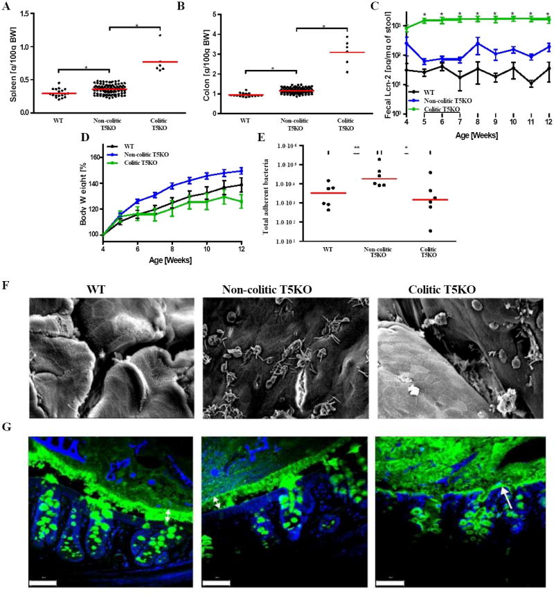
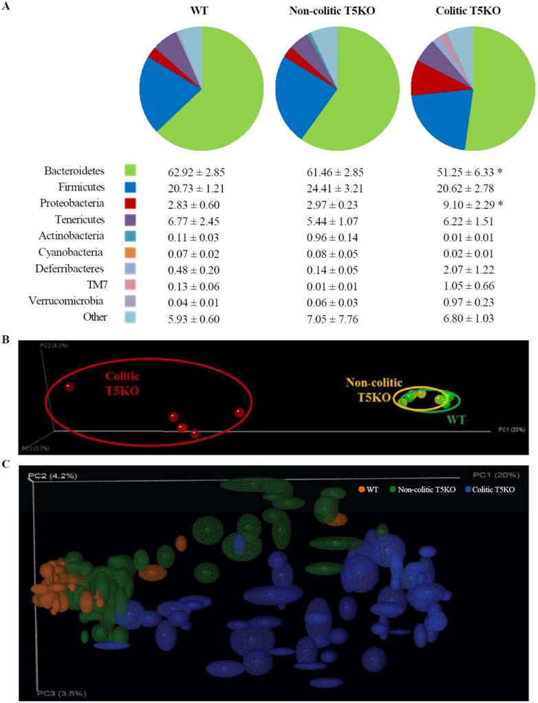
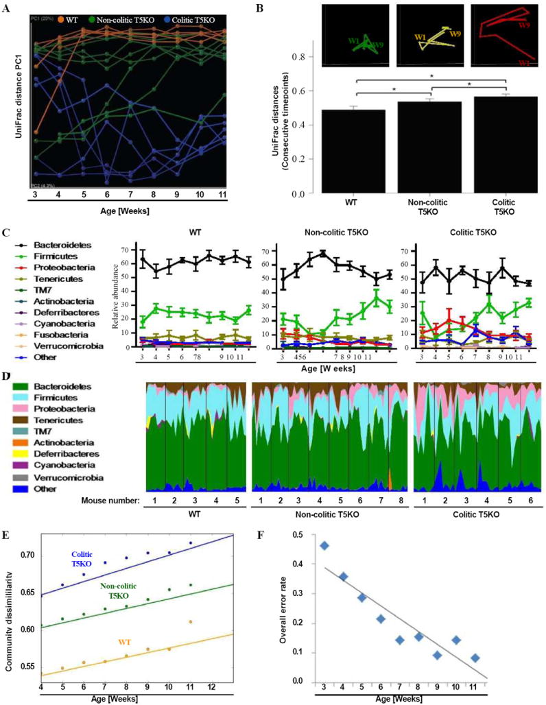
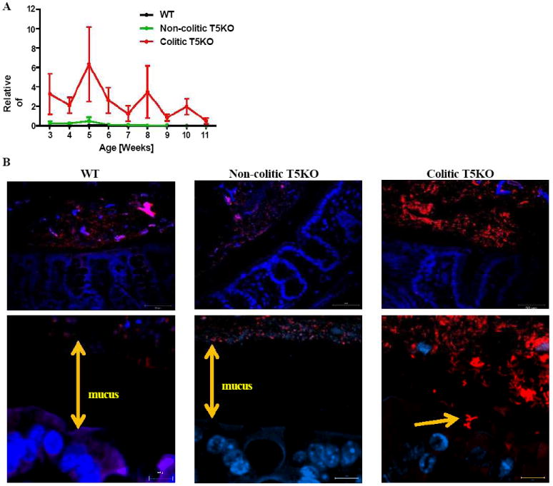
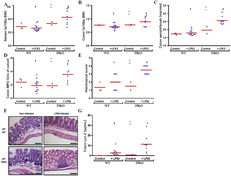
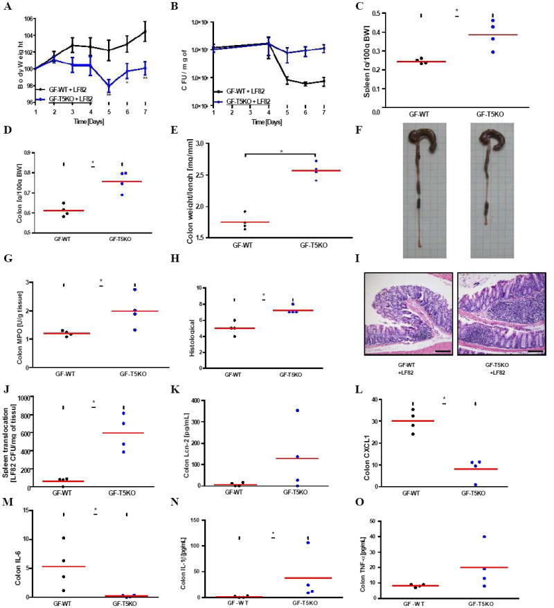
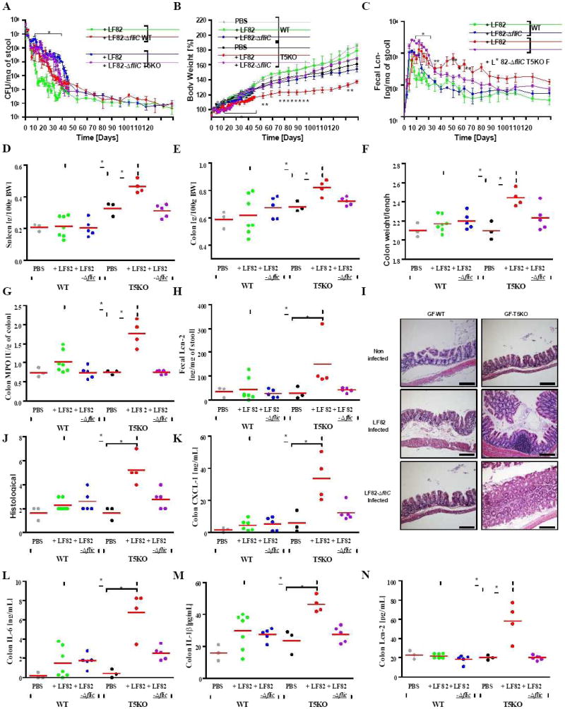
Colitis results from breakdown of homeostasis between intestinal microbiota and the mucosal immune system, with both environmental and genetic influencing factors. Flagellin receptor TLR5-deficient mice (T5KO) display elevated intestinal proinflammatory gene expression and colitis with incomplete penetrance, providing a genetically sensitized system to study the contribution of microbiota to driving colitis. Both colitic and noncolitic T5KO exhibited transiently unstable microbiotas, with lasting differences in colitic T5KO, while their noncolitic siblings stabilized their microbiotas to resemble wild-type mice. Transient high levels of proteobacteria, especially enterobacteria species including E. coli, observed in close proximity to the gut epithelium were a striking feature of colitic microbiota. A Crohn's disease-associated E. coli strain induced chronic colitis in T5KO, which persisted well after the exogenously introduced bacterial species had been eliminated. Thus, an innate immune deficiency can result in unstable gut microbiota associated with low-grade inflammation, and harboring proteobacteria can drive and/or instigate chronic colitis.


Figures

Figure 1
Figure 1. Development of spontaneous inflammation and microbial dysbiosis in T5KO mice
Wild-type (n=18) and T5KO mice (n=110) were housed for 12 weeks in the animal facility to track spontaneous colitic mice (defined as described in Methods). (A) Following euthanasia, spleen was isolated and mass measured. (B) Colon mass. (C) Stool was collected weekly after weaning and diluted in 500 μL of PBS. Then, supernatant was assayed for lipocalin-2 (Lcn-2) expression by ELISA. (D) Body mass was monitored weekly from week 4 to week 12. (E) Colon was washed carefully with PBS to remove any stool and bacterial DNA was isolated. Total adherent bacteria was measured by quantitative PCR analysis using universal 16S rRNA primers. (F) Representative electron microscopy observation of colon (magnification: 1500X). (G) Muc 2 mucin immunostaining - green. Nuclei were stained using DAPI - blue (Scale bar = 20 μM). The data on panels E-G is representative of 2 independent experiments.* p<0.05.
Figure 2
Figure 2. End point microbiota composition in WT, non-colitic and colitic T5KO mice
Wild-type, non-colitic and colitic T5KO mice (n=6-8 mice per group) were euthanized at 12-week old. Cecal contents were collected and microbiota composition was analyzed via 16S rRNA analysis. (A) Relative abundance of phyla in cecal bacteria. Table provides mean for each phyla as the percentage of total sequences analyzed. (B) Mouse cecal bacterial communities were clustered using principal coordinates analysis (PCoA) of the UniFrac unweighted distance matrix. PC1, PC2 and PC3 are plotted. The percentage of the variation explained by the plotted principal coordinates is indicated in the axis labels. Results are from an analysis of 5-6 mice per group. (C) Jackknifing PCoA plot of cecal and fecal samples from all time points. Point locations are the average location of 10 jackknife replicates using 1000 random sequences per sample and ellipses show the confidence based on these randomizations. Analysis was done by ANOVA and statistical significance (P<0.01) is denoted by asterisk (*).
Figure 3
Figure 3. Increased volatility in microbiota of T5KO mice
Stool from wild-type, non-colitic and colitic T5KO mice (n=5-8 mice per group) were collected weekly for 9 weeks after weaning (from 3-week to 11-week old). Stool microbiota composition was analyzed via 16S rRNA analysis. (A) Mouse cecal bacterial communities were clustered using principal coordinates analysis (PCoA) of the UniFrac unweighted distance matrix. PC1 is plotted for each time point (from 3-week to 11-week old). The time is expressed on the x-axis and the percentage of the variation explained by the plotted principal coordinates is indicated in the y-axis labels. Results are from an analysis of 5-8 mice per group (samples generating less than 1000 sequences were removed from analysis). (B) After clustering of mouse cecal bacterial communities using principal coordinates analysis (PCoA) of the UniFrac unweighted distance matrix, a representative mouse has been used to illustrate the time point evolution of the microbiota (top panel). The average of the UniFrac unweighted distance for each category (WT, non-colitic and colitic T5KO) between consecutive time points has been calculated (bottom panel). (C) Relative abundance of phyla in stool bacteria from WT (left panel), non-colitic T5KO (middle panel) and colitic T5KO (right panel) mouse group. (D) Relative abundance of phyla over time (from 3-week to 11-week old) in stool bacteria from individual WT (left panel), non-colitic T5KO (middle panel) and colitic T5KO (right panel) mice. (E) Semivariogram plot of community dissimilarity (UniFrac, y axis) vs. days dissimilarity (Euclidean, x axis). (F) Overall misclassification error rates using 9 families at each week. Analysis was done by ANOVA and statistical significance (P<0.01) is denoted by asterisk (*). Related materials can be found in supplementary figure 1.
Figure 4
Figure 4. Colitic T5KO mice harbored abnormal amount of Enterobacteria
(A) Stool from wild-type, non-colitic and colitic T5KO mice (n=6-8 mice per group) were collected weekly for 9 weeks after weaning (from 3-week to 11-week old). Stool microbiota composition was analyzed via 16S rRNA analysis to determine the relative abundance of Enterobacteria in stool from WT, non-colitic T5KO and colitic T5KO mouse group (B) Microscopic pictures of colon after FISH using an Alexa 555 conjugated Enterobacteria specific probe (red) and nuclei in blue after DAPI staining (magnification: top-40X, bottom-100X). Related materials can be found in supplementary figure 2.
Figure 5
Figure 5. Mono-association of Crohn’s disease associated Adherent-Invasive E. coli (AIEC) strain LF82 infection increased intestinal inflammation in T5KO mice
(A) Following euthanasia, spleen was isolated and mass measured. (B) Colon mass. (C) Ratio between colon weight and colon length. (D) Colon MPO activity. (E-F) Histological score and representative H&E stained colon (magnification, 100×). (G) Colon was cultured for 24h, at which time supernatant was assayed for Lcn-2 by ELISA. * p<0.05. Related materials can be found in supplementary figure 3.
Figure 6
Figure 6. Germ-free T5KO mice are highly susceptibility to early AIEC LF82 infection
Germ-free wild-type and T5KO mice (n=4 mice per group) were orally infected with 107 flagellate AIEC LF82 bacteria. (A) Body mass was monitored daily during the treatment. (B) Numeration of AIEC LF82 present in the WT or T5KO mouse stool from day 1 to day 7 post infection. (C) Following euthanasia, spleen was isolated and mass measured. (D) Colon mass. (E) Ratio between colon weight and colon length. (F) Gross picture of colon. (G) Colon MPO activity. (H-I) Histological score and representative H&E stained colon (magnification, 100×). (J) Bacterial translocation by numbering AIEC LF82 CFU present in the spleen at day 7 post infection. (K-O) Colon was cultured for 24h, at which time supernatant was assayed for Lcn-2 (K) and several pro-inflammatory cytokines, namely CXCL1 (L), IL-6 (M), IL-1β (N) and TNF-α (O), by ELISA. * p<0.05. Related materials can be found in supplementary figure 4.
Figure 7
Figure 7. Transient AIEC LF82 infection resulted in a chronic basal inflammation in germ-free T5KO mice
Germ-free wild-type and T5KO mice (n=3-7 mice per group) were orally infected with 107 flagellate AIEC LF82 bacteria. As control, uninfected germ-free WT or T5KO mice were transferred to a sterile cage without treatment. (A) Numeration of AIEC LF82 present in the WT or T5KO mouse stool. (B) Body mass was monitored daily from day 0 to day 35 post infection and then weekly from day 42 to day 119 post infection. (C) Stool was collected daily (D0 to D35) or weekly (D42 to D119) after AIEC LF82 infection and diluted in 500 μL of PBS. Then, supernatant was assayed for Lcn-2 expression by ELISA. (D) Following euthanasia, spleen was isolated and mass measured. (E) Colon mass. (F) Ratio between colon weight and colon length. (G) Colon MPO activity. (H) Fecal Lcn-2 was assayed by ELISA at the end of the experiment. (I-J) Histological score and representative H&E stained colon (magnification, 100×). (K-N) Colon was cultured for 24h, at which time supernatant was assayed for several pro-inflammatory cytokines, namely CXCL1 (K), IL-6 (L), IL-1β (M), and Lcn-2 (N) by ELISA. * p<0.05. Related materials can be found in supplementary figure 5.

References

    1. Antunes LC, Finlay BB. A comparative analysis of the effect of antibiotic treatment and enteric infection on intestinal homeostasis. Gut Microbes. 2011;2:105–108. - PubMed
    1. Barnich N, Boudeau J, Claret L, Darfeuille-Michaud A. Regulatory and functional co-operation of flagella and type 1 pili in adhesive and invasive abilities of AIEC strain LF82 isolated from a patient with Crohn’s disease. Mol Microbiol. 2003;48:781–794. - PubMed
    1. Barthel M, Hapfelmeier S, Quintanilla-Martinez L, Kremer M, Rohde M, Hogardt M, Pfeffer K, Russmann H, Hardt WD. Pretreatment of mice with streptomycin provides a Salmonella enterica serovar Typhimurium colitis model that allows analysis of both pathogen and host. Infect Immun. 2003;71:2839–2858. - PMC - PubMed
    1. Carvalho FA, Aitken JD, Gewirtz AT, Vijay-Kumar M. TLR5 activation induces secretory interleukin-1 receptor antagonist (sIL-1Ra) and reduces inflammasome-associated tissue damage. Mucosal Immunol. 2011;4:102–111. - PMC - PubMed
    1. Carvalho FA, Barnich N, Sauvanet P, Darcha C, Gelot A, Darfeuille-Michaud A. Crohn’s disease-associated Escherichia coli LF82 aggravates colitis in injured mouse colon via signaling by flagellin. Inflamm Bowel Dis. 2008;14:1051–1060. - PubMed

Publication types

Substances

LinkOut - more resources