. 2012 Oct;141(1):8-14.
doi: 10.1016/j.schres.2012.07.007. Epub 2012 Aug 3.

A broken filter: prefrontal functional connectivity abnormalities in schizophrenia during working memory interference

Affiliations

A broken filter: prefrontal functional connectivity abnormalities in schizophrenia during working memory interference

Alan Anticevic et al. Schizophr Res. 2012 Oct.

Abstract

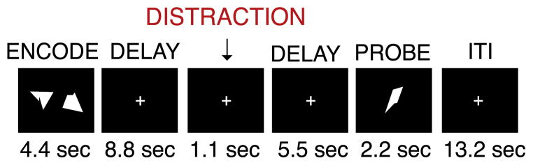
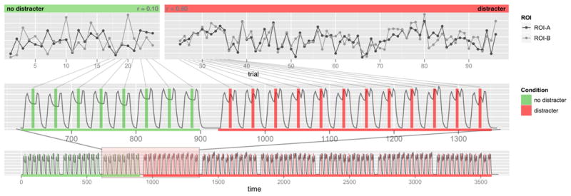
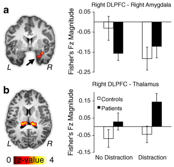
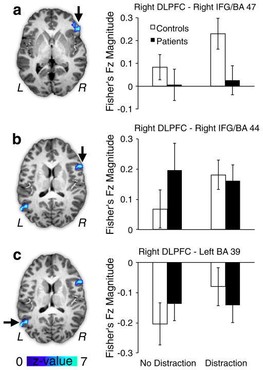
Characterizing working memory (WM) abnormalities represents a fundamental challenge in schizophrenia research given the impact of cognitive deficits on life outcome in patients. In prior work we demonstrated that dorsolateral prefrontal cortex (DLPFC) activation was related to successful distracter resistance during WM in healthy controls, but not in schizophrenia. Although understanding the impact of regional functional deficits is critical, functional connectivity abnormalities among nodes within WM networks may constitute a final common pathway for WM impairment. Therefore, this study tested the hypothesis that schizophrenia is associated with functional connectivity abnormalities within DLPFC networks during distraction conditions in WM. 28 patients and 24 controls completed a delayed non-verbal WM task that included transient visual distraction during the WM maintenance phase. We computed DLPFC whole-brain task-based functional connectivity (tb-fcMRI) specifically during the maintenance phase in the presence or absence of distraction. Results revealed that patients failed to modulate tb-fcMRI during distracter presentation in both cortical and sub-cortical regions. Specifically, controls demonstrated reductions in tb-fcMRI between DLPFC and the extended amygdala when distraction was present. Conversely, patients failed to demonstrate a change in coupling with the amygdala, but showed greater connectivity with medio-dorsal thalamus. While controls showed more positive coupling between DLPFC and other prefrontal cortical regions during distracter presentation, patients failed to exhibit such a modulation. Taken together, these findings support the notion that observed distracter resistance deficit involves a breakdown in coupling between DLPFC and distributed regions, encompassing both subcortical (thalamic/limbic) and control region connectivity.


Conflict of interest statement

Conflict of Interest

John H. Krystal, MD 2012 financial disclosure.

Note: the individual consultant agreements listed below are less than $10,000 per year.

Consultant

  1. Aisling Capital, LLC

  2. Astellas Pharma Global Development, Inc.

  3. AstraZeneca Pharmaceuticals

  4. Biocortech

  5. Brintnall & Nicolini, Inc.

  6. Easton Associates

  7. Gilead Sciences, Inc.

  8. GlaxoSmithKline

  9. Janssen Pharmaceuticals

  10. Lundbeck Research USA

  11. Medivation, Inc.

  12. Merz Pharmaceuticals

  13. MK Medical Communications

  14. F. Hoffmann-La Roche Ltd

  15. Sage Therapeutics, Inc.

  16. SK Holdings Co., Ltd

  17. Sunovion Pharmaceuticals, Inc.

  18. Takeda Industries

  19. Teva Pharmaceutical Industries, Ltd.

Scientific Advisory Board

  1. Abbott Laboratories

  2. Bristol-Myers Squibb

  3. CHDI Foundation, Inc.

  4. Eisai, Inc.

  5. Eli Lilly and Co.

  6. Forest Laboratories, Inc.

  7. Lohocla Research Corporation

  8. Mnemosyne Pharmaceuticals, Inc.

  9. Naurex, Inc.

  10. Pfizer Pharmaceuticals

  11. Shire Pharmaceuticals

  12. StratNeuro Research Program at Karolinska Institute (International Advisory Board)

Board of Directors

  1. Coalition for Translational Research in Alcohol and Substance Use Disorders

President

  1. American College of Neuropsychopharmacology

Income greater than $10,000

Editorial Board

  1. Editor - Biological Psychiatry

Employment

  1. Yale University School of Medicine

  2. VA CT Healthcare System

Patents and inventions

  1. Seibyl JP, Krystal JH, Charney DS. Dopamine and noradrenergic reuptake inhibitors in treatment of schizophrenia. Patent #:5,447,948.September 5, 1995

  2. I am a co-inventor with Dr. Gerard Sanacora on a filed patent application by Yale University related to targeting the glutamatergic system for the treatment of neuropsychiatric disorders (PCTWO06108055A1).

  3. Intranasal administration of ketamine to treat depression (pending)

Figures

Fig. 1
Fig. 1
Task Design. Overall task design is shown. For the purposes of the present investigation we collapsed across different distracter conditions (see Method) since patients were more distracted than controls across all distracter types irrespective of distracter condition (Anticevic et al., 2011c). Complete details regarding the task were described previously (Anticevic et al., 2011b, 2011c). We also provide additional task details and considerations in the Supplement.
Fig. 2
Fig. 2
tb-fcMRI time-point selection approach. We illustrate the tb-fcMRI analysis strategy using the slow event-related design. This approach closely follows our previously published work (Anticevic et al., 2010b). The bottom panel shows the time series across the entire experiment. The initial time series marked in green indicates trials with no distraction, followed by trials with distraction marked in red. The middle panel focuses on a sub-set of the trials to more closely illustrate the time-point selection strategy. The vertical bars mark the corresponding ‘middle’ portion of each trial where activity is sampled by averaging across two frames following the onset of distraction. The top panel illustrates how these frames are concatenated into a time-series representing distracter-related signal across all trials. All tb-fcMRI analyses are performed on these extracted time courses, which reflect variation in peak response – as indicated by obtained correlation coefficients shown in corners of each top panel. This analytic strategy largely circumvents the concern that correlations are being driven by overall task response.
Fig. 3
Fig. 3
Subcortical regions showing significant tb-fcMRI group differences with right DLPFC following WM interference. All regions exhibited a significant Diagnosis (patients vs. controls) x Distraction (no distraction vs. distraction) interaction at the whole-brain level. (a) tb-fcMRI is shown between right DLPFC and right limbic cortex, proximal to the right amygdala (x=29, y=−3, z=−20). Controls (white bars) showed more negative coupling between right DLPFC and right amygdala, whereas patients (black bars) failed to exhibit such modulation. (b) tb-fcMRI is shown between right DLPFC and bilateral dorsal thalamic region (right: x=15, y=−26, z=15; left: x=12, y=−24, z=14). Patients (black bars) exhibited increases in right DLPFC-thalamic connectivity specifically following WM interference, whereas for controls this connectivity was attenuated (white bars). Error bars reflect +/− 1 standard error of the mean. For a vertical scatterplot showing the full distribution of all participants please see Supplement.
Fig. 4
Fig. 4
Cortical regions showing significant tb-fcMRI group differences with right DLPFC following WM interference. As in Fig. 1, all regions exhibited a significant Diagnosis (patients vs. controls)×Distraction (no distraction vs. distraction) interaction at the whole-brain level. (a) tb-fcMRI is shown between right DLPFC and right inferior frontal gyrus (IFG), corresponding to Brodmann’s area 47 (x=52, y=28, z=0). Patients (black bars) failed to show an increase in right DLPFC-IFG connectivity, whereas controls (white bars) showed a clear increase in coupling following WM interference. (b) tb-fcMRI is shown between right DLPFC and a more inferior portion of the right IFG, proximal to Brodmann’s area 44 (x=53, y=11, z=15). Again, controls showed an increase in positive coupling following interference, whereas patients failed to exhibit this modulation. (c) tb-fcMRI is shown between right DLPFC and left parietal cortex proximal to Brodmann’s area 39 (x=−53, y=−60, z=18). Controls showed a reduction of negative coupling in response to distraction, but patients failed to show a modulation of DLPFC-parietal coupling. Error bars reflect +/− 1 standard error of the mean. For a vertical scatterplot showing the full distribution of all participants please see Supplement.

References

    1. Andreasen NC. The scale for the assessment of negative symptoms (SANS) University of Iowa, Iowa City; 1983a.
    1. Andreasen NC. The scale for the assessment of positive symptoms (SAPS) University of Iowa, Iowa City; 1983b.
    1. Anticevic A, Repovs G, Barch DM. Resisting Emotional Interference: Brain Regions Facilitating Working Memory Performance During Negative Distraction. Cogn Affect Behav Neurosci. 2010a;10 (2):159–173. - PMC - PubMed
    1. Anticevic A, Repovs G, Shulman GL, Barch DM. When less is more: TPJ and default network deactivation during encoding predicts working memory performance. NeuroImage. 2010b;49:2638–2648. - PMC - PubMed
    1. Anticevic A, Repovs G, Corlett PR, Barch DM. Negative and Non-emotional Interference with Visual Working Memory in Schizophrenia. Biol Psychiatry. 2011a;70 (12):1159–1168. - PubMed

Publication types

MeSH terms