. 2012 Aug 14;23(2):292-304.
doi: 10.1016/j.devcel.2012.07.002. Epub 2012 Aug 2.

Sizzled-tolloid interactions maintain foregut progenitors by regulating fibronectin-dependent BMP signaling

Affiliations

Sizzled-tolloid interactions maintain foregut progenitors by regulating fibronectin-dependent BMP signaling

Alan P Kenny et al. Dev Cell. .

Abstract

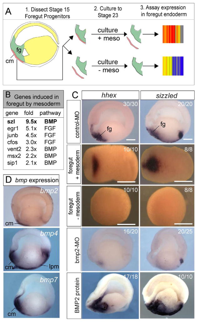
The liver, pancreas, and lungs are induced from endoderm progenitors by a series of dynamic growth factor signals from the mesoderm, but how the temporal-spatial activity of these signals is controlled is poorly understood. We have identified an extracellular regulatory loop required for robust bone morphogenetic protein (BMP) signaling in the Xenopus foregut. We show that BMP signaling is required to maintain foregut progenitors and induce expression of the secreted frizzled related protein Sizzled (Szl) and the extracellular metalloprotease Tolloid-like 1 (Tll1). Szl negatively regulates Tll activity to control deposition of a fibronectin (FN) matrix between the mesoderm and endoderm, which is required to maintain BMP signaling. Foregut-specific Szl depletion results in a loss of the FN matrix and failure to maintain robust pSmad1 levels, causing a loss of foregut gene expression and organ agenesis. These results have implications for BMP signaling in diverse contexts and the differentiation of foregut tissue from stem cells.


Figures

Figure 1
Figure 1. Mesodermal BMP2 signals maintain foregut progenitors and induce szl
(A) Strategy to identify genes induced in the foregut progenitors (fg; green) by the precardiac mesoderm (cm; red). (B) BMP and FGF pathway genes identified by microarray. (C) In situ hybridization to bisected stage 20 embryos (anterior left) and isolated ventral explants cultured from stage 15 to 23 with or without mesoderm confirms that szl and hhex expression requires mesodermal signals. Injection of cont-MO, bmp2-MO, or BMP2 protein demonstrates that szl and hhex expression are regulated by BMP signaling. (D) Bisected stage 20 embryos (anterior left) showing bmp2, bmp4 and bmp7 expression in the precardiac (cm) and the ventral lateral plate mesoderm (lpm). See also Figure S1.
Figure 2
Figure 2. Sizzled is required for foregut organogenesis
(A–B) Lineage labeling by a fluorescent dextran (FLDX) co-injected with a szl-MO (20ng) into the C1/D1 cells of the 16-cell-stage embryo targets the dorsal-anterior mesendoderm at gastrula stage 10.5 (A), which gives rise to the ventral foregut progenitors at stage 20 (B). (C–D) A comparison to szl expression shows that these injections avoid the szl-expressing ventral mesoderm at stage 10.5, but targets foregut szl at stage 20. d; dorsal, v; ventral, a; anterior, p; posterior. (E–P) In situ hybridization of control and szl-MO injected embryos demonstrates that Szl is required for foregut organogenesis. At stage 20 szl-MO embryos failed to maintain hhex expression, while nxk2.5 in the pre-cardiac mesoderm was unaffected (E–H). Analysis of pdx1 (pancreas), nkx2.1 (lung/thyroid), hhex (liver/thyroid) and tnlc (heart) at stage 35 demonstrates that Szl is required for foregut organ specification. (Q and R) H&E staining of stage 37 embryos reveals a reduction in foregut size (dashed outline) and cell sloughed into the pharynx of szl-MO. (S and T) Isolated gut tubes at stage 42 show that szl-MO lack foregut organ buds (lu; lung, liver lv; liver, p; pancreas). See also Figure S2.
Figure 3
Figure 3. Szl depletion results in foregut progenitor apoptosis
(A) Szl-depletion causes increased foregut cell apoptosis. Mean number of activated caspase-3 cells ± SEM in the foreguts of stage 20 embryos injected with; cont-MO (20 ng), szl-MO (15 ng) and/or szl-DNA (500 pg), from 3 independent injection experiments. In student T-tests p<0.05 *compared to cont-MO and ** compared to szl-MO. (B) Apoptosis was specific to the foregut. The szl-MO (20ng) was injected in all cells of 4-cell stage embryos and the mean number of activated caspase-3 positive cells ± SEM in the foregut, hindgut and ectoderm was scored from five stage 20 embryos. *p<0.05 comparing cont-MO to szl-MO, ns= not significant. (C–D) Cell death cannot account for loss of foregut identity. szl-MO injected embryos treated at stage 12 with one of three different caspase inhibitors (n>10 per inhibitor). In each case szl-MO embryos still exhibit reduced hhex and for1 (C) even when cell death was rescued (D). A representative experiment is shown.
Figure 4
Figure 4. pSmad1 dynamics in the Xenopus foregut
(A) Diagram of a stage 20 Xenopus embryo section showing the foregut (fg) region in green that was isolated for western blots and imaged by confocal. (B) Immunoblot of pSmad1/5/8, total Smad1 and Tubulin levels in foregut explants at the indicated developmental stages, with the pSmad1/total Smad1 ratio quantitated above. (C) Immunostaining of pSmad1/5/8 (red) and nuclei (green) in the foregut of bisected embryos at the indicated stage shows spatially and temporally dynamic BMP signaling in the foregut. White dashed lines indicate the boundaries between the endoderm (end), mesoderm (m) and ectoderm (ect).
Figure 5
Figure 5. Szl is required to maintain robust BMP signaling in the foregut
(A) Western blot of pSmad1/5/8 and total Smad1 in foregut explants from cont-MO and szl-MO embryos at different stages shows reduced BMP signaling in Szl-morphants. (B) Szl-depleted embryos exhibit reduced pSmad1 signaling in the foregut. 50μm confocal Z-projections of pSmad1/5/8 (green) and nuclei (red) in stage 20 sibling embryos injected with either; cont-MO (20ng), szl-MO (15 ng), bmp2-MO (20 ng) or treated with BMP-receptor inhibitor LDN193189 (40 μM). Yellow dashed line indicates the mesoderm-endoderm boundary. Dashed white lines in nuclear channel of the cont-MO illustrate the sub-region of the foregut, based on number of nuclei from the mesoderm. See also Figure S3. (C) Szl and Bmp2 depleted embryos have reduced pSmad1 levels in the deep foregut endoderm. Mean nuclear/cytoplasmic ratio of pSmad1 immunostaining intensity ±SEM in foregut cells at different distances from the mesoderm, quantitated from 80 μm Z-projections from 4 sibling embryos/condition. *Significantly different from szl-MO, p<0.05. (D) Expression of bmp2, bmp4/7 and BMP-target genes szl, vent1/2, and hhex were down regulated by injection of the szl-MO (15 ng) and rescued by subsequent injection of recombinant BMP2 protein (4 ng) into the foregut at stage 12. (E) Mean number of activated caspase-3 positive foregut cells ± SEM (n=5 embryos/condition), * p<0.01 in Student T-tests.
Figure 6
Figure 6. Knockdown of Tll proteases rescues the szl-MO phenotype
(A–D) Expression of szl, tll1, bmp1 and chordin in stage 18 embryos. (E–H) tll1 expression in embryos injected in the C1/D1 cells with either; control-MO (20ng), szl-MO (15 ng), bmp2-MO (20 ng) or treated with BMP-receptor inhibitor LDN193189 (40 μM). (I–T) Knockdown of Tll1 and Bmp1 rescues the Szl morphant phenotype. tll1-MOs and bmp1-MOs (15 ng each) were injected into C1/D1 cells with or without szl-MO (15 ng) and assayed for hhex (I–L) and vent1/2 (M–P) at stage 20 and the liver marker for1 at stage 35 (Q–T). See also Figure S4.
Figure 7
Figure 7. Loss of FN matrix in szl-MO
(A) FN and β1-integrin immunostaining (10 μm confocal slice) of cont-MO and szl-MO injected foreguts at stage 18–20 shows that the FN matrix between the endoderm (end) and mesoderm (mes) (yellow arrows) is absent in szl-MO (red arrows). Yellow dashed lines outline the precardiac lateral plate mesoderm (B) Western blots of foregut explants showing no obvious degradation of FN or β1-Integrin in Szl morphants. The relative FN/tubulin and Integrin/tubulin levels in the szl-MO compared to the cont-MO, which was set to 1.0 is indicated. See also Figure S5.
Figure 8
Figure 8. Szl-Tll regulate FN matrix assembly, which is required to maintain BMP signaling
(A) Embryos injected with either; cont-MO (20ng), szl-MO (15 ng), cont-MO + bmp1-MO + tll1-MO (15 ng each), szl-MO + bmp1-MO + tll1-MO (15 ng each), fn-MOs (15 ng) or treated with BMP-receptor inhibitor LDN193189 (40 μM). Confocal FN immumostaining (10 μm slice) shows FN fibrils (yellow arrow heads) between the foregut endoderm (end) and mesoderm (m), which are disrupted in szl-MO and fn-MO injected embryos (red arrow heads). Confocal immunostaining of pSmad1 (green) and nuclei (red) in the foregut endoderm (50 μm Z-projection 4–9 nuclei from the mesoderm) and in situ for bmp2, bmp4/7 and hhex indicate that BMP signaling was reduced in szl-MO, fn-MO, and LDN treated embryos. Knockdown of bmp1/tll1 rescues the FN matrix, pSmad1 levels and BMP-responsive gene expression in Szl morphants. See also Figure S6. (B) Mean nuclear/cytoplasmic pSmad1 immunostaining intensity ±SEM in foregut cells located 4–9 nuclei from the mesoderm, quantitated from 80 μm Z-projection from 4 sibling embryos/condition. Significantly different from contMO in T-test: *p<0.05, **LS mean difference >0.3, p<0.01, ns = not significant. (C) Mean number of activated caspase-3 foregut cells ±SEM. *p<0.05 compared to cont-MO.

Similar articles

Cited by

References

    1. Blitz IL, Shimmi O, Wunnenberg-Stapleton K, O’Connor MB, Cho KW. Is chordin a long-range- or short-range-acting factor? Roles for BMP1-related metalloproteases in chordin and BMP4 autofeedback loop regulation. Dev Biol. 2000;223:120–138. - PubMed
    1. Bovolenta P, Esteve P, Ruiz JM, Cisneros E, Lopez-Rios J. Beyond Wnt inhibition: new functions of secreted Frizzled-related proteins in development and disease. J Cell Sci. 2008;121:737–746. - PubMed
    1. Chung WS, Andersson O, Row R, Kimelman D, Stainier DY. Suppression of Alk8-mediated Bmp signaling cell-autonomously induces pancreatic beta-cells in zebrafish. Proc Natl Acad Sci U S A. 2010;107:1142–1147. - PMC - PubMed
    1. Chung WS, Shin CH, Stainier DY. Bmp2 signaling regulates the hepatic versus pancreatic fate decision. Dev Cell. 2008;15:738–748. - PMC - PubMed
    1. Collavin L, Kirschner MW. The secreted Frizzled-related protein Sizzled functions as a negative feedback regulator of extreme ventral mesoderm. Development. 2003;130:805–816. - PubMed

Publication types

MeSH terms

Associated data