. 2012 Oct;181(4):1306-15.
doi: 10.1016/j.ajpath.2012.06.016. Epub 2012 Aug 3.

p53 mediates TNF-induced epithelial cell apoptosis in IBD

Affiliations

p53 mediates TNF-induced epithelial cell apoptosis in IBD

Tatiana Goretsky et al. Am J Pathol. 2012 Oct.

Erratum in

  • Am J Pathol. 2013 Feb;182(2):610

Abstract

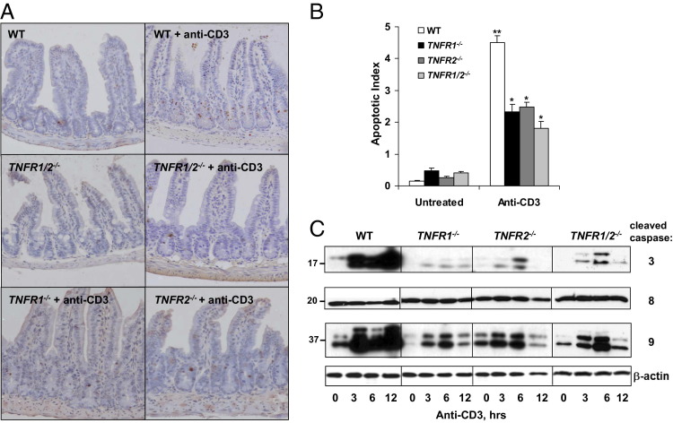
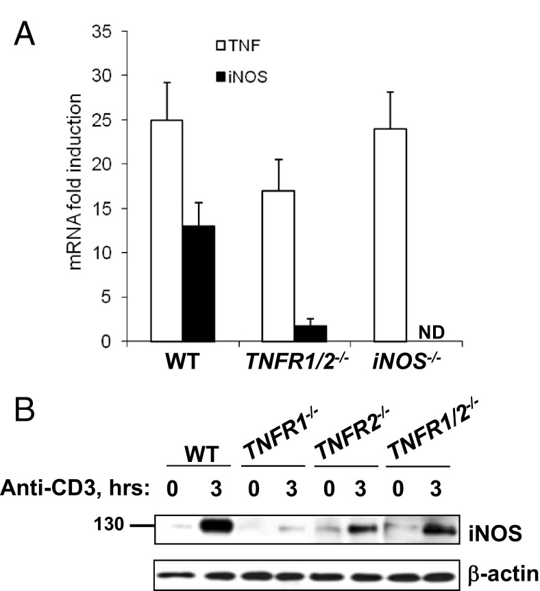
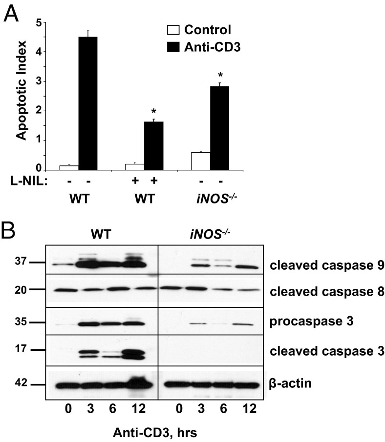
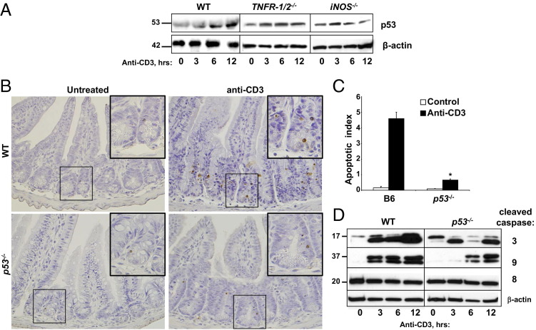
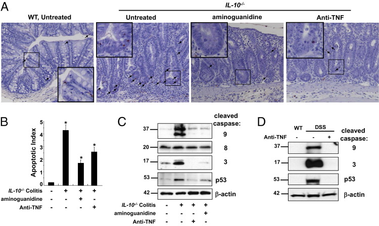
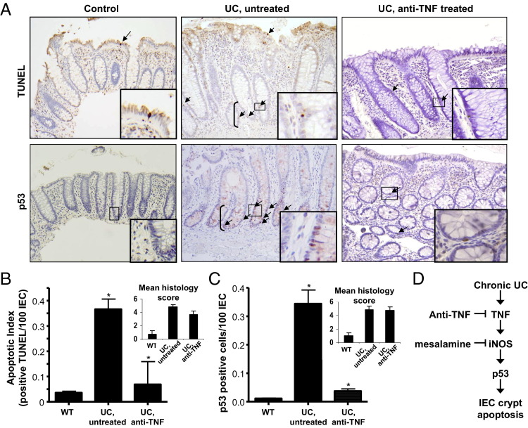
Chronic ulcerative colitis (CUC) is characterized by increased intestinal epithelial cell (IEC) apoptosis associated with elevated tumor necrosis factor (TNF), inducible nitric oxide synthase (iNOS), and p53. We previously showed that p53 is increased in crypt IECs in human colitis and is needed for IEC apoptosis in chronic dextran sulfate sodium-colitis. Herein, we examined the roles of TNF and iNOS in regulating p53-induced IEC apoptosis in CUC. The IEC TUNEL staining, caspases 3, 8, and 9, and p53 protein levels, induced by anti-CD3 monoclonal antibody (mAb) activation of T cells, were markedly reduced in TNF receptor 1 and 2 gene knockout mice. Induction of IEC apoptosis correlated with increased p53, which was attenuated in iNOS(-/-) mice. IEC p53 levels and apoptosis were reduced in IL-10(-/-) colitic mice treated with neutralizing TNF mAb and the iNOS inhibitor, aminoguanidine, further suggesting that TNF and iNOS are upstream of p53 during colitis-induced IEC apoptosis. IEC apoptosis and p53 levels were assessed in control versus untreated or anti-TNF-treated CUC patients with equivalent levels of inflammation. Data indicated that IEC apoptosis and p53 levels were clearly higher in untreated CUC but markedly reduced in patients treated with anti-TNF mAb. Therefore, TNF-induced iNOS activates a p53-dependent pathway of IEC apoptosis in CUC. The inhibition of IEC apoptosis may be an important mechanism for mucosal healing in anti-TNF-treated CUC patients.


Figures

Figure 1
Figure 1
TNFR1 and TNFR2 signaling induces TNF-mediated crypt cell apoptosis. WT, TNFR1/, TNFR2/, and TNFR1/2/ mice are injected with control or anti-CD3 antibody and sacrificed at the indicated time points. A: TUNEL staining of SB crypts from mice sacrificed 24 hours after anti-CD3 mAb injection. B: The apoptotic index is determined based on counting TUNEL-positive cells. P < 0.0001 compared with WT stimulated mice; ⁎⁎P < 0.0001 for anti-CD3-treated WT mice compared with untreated WT mice. C: WB of activated caspases 3, 8, and 9 from CD45-negative (data not shown) IEC lysates resolved by SDS-PAGE. β-Actin is used as a loading control.
Figure 2
Figure 2
iNOS mediates T-cell–induced crypt cell apoptosis downstream of TNFR1 and TNFR2. A: Fold induction of iNOS and TNF mRNA expression 3 hours after anti-CD3 injection determined by real-time RT-PCR from SB tissue from WT, TNFR1/2−/−, and iNOS/ mice. B: WB analysis of iNOS protein expression in the indicated mice before, or 3 hours after, T-cell activation. β-Actin is used as a loading control. ND, not determined.
Figure 3
Figure 3
iNOS is required for IEC apoptosis. A: Mice are sacrificed 24 hours after control or anti-CD3 mAb injection, and apoptosis is quantified in SB sections from WT mice, with or without L-NIL treatment, and from iNOS/ mice. Apoptotic indexes are calculated based on counting TUNEL-positive cells. P < 0.001 compared with anti-CD3-treated WT mice. B: Activation of caspases 3, 8, and 9 is assessed by WB for the cleaved forms of the indicated protein in WT and iNOS/ mice at 0, 3, 6, or 12 hours after T-cell activation. Procaspase 3 is also shown, and β-actin is used as a loading control.
Figure 4
Figure 4
p53 Plays a role in T-cell–induced crypt cell apoptosis downstream of TNFR1, TNFR2, and iNOS. A: WB analysis of p53 protein expression and stabilization in SB IECs from WT, TNFR1/2/, and iNOS/ mice. B: Apoptosis is assessed in the SB crypts and villi in p53/ mice by TUNEL staining 24 hours after stimulation with anti-CD3 mAb injection relative to WT mice. Insets: Areas magnified (original magnification, ×40) are demarcated by boxes in the ×10 picture. C: Results are quantified by counting TUNEL-positive cells to determine the apoptotic index. P < 0.0001. D: Caspase activation is determined by WB analysis at the indicated times after anti-CD3 injection. β-Actin is used as a loading control.
Figure 5
Figure 5
Inhibitors of TNF and iNOS decrease colitis-induced IEC apoptosis. Colitis is induced in IL-10/ mice with piroxicam, and apoptosis is assessed at day 28. Mice are treated with aminoguanidine to block iNOS or anti-TNF mAb to inhibit TNF. Colonic IECs undergoing apoptosis are identified by TUNEL staining (A, arrows), and the apoptotic index is calculated based on cell counting of TUNEL-positive cells (B). P < 0.004 compared with colitic mice; ⁎⁎P < 0.0001 compared with noncolitic mice. High-power magnification (×40) is shown in the small boxes in the low-magnification (×10) pictures. C: WB analysis of colon IEC whole cell lysates for caspase 3, 8, and 9 activation and for p53 levels. Chronic colitis is also induced in WT mice after three cycles of DSS. D: Mice are either left untreated or treated with anti-TNF mAb, and levels of p53 and cleaved caspases 3 and 9 are analyzed. β-Actin is used as a loading control.
Figure 6
Figure 6
Apoptosis and p53 expression are induced by TNF in human UC. Colon tissue biopsy specimens from normal control, UC untreated, and UC anti-TNF–treated patients are analyzed for p53 and TUNEL staining (A, arrows) and then quantified by counting positive cells per 100 IECs (B and C). Insets: Areas magnified (×40) are demarcated by boxes. Untreated and anti-TNF–treated UC patients selected for study are inflamed, as indicated by mean histological scores (B and C). *P < 0.01 (B), P < 0.0001 (C) for UC untreated compared to anti-TNF–treated patients. The proposed paradigm for IEC crypt apoptosis, in which chronic inflammation from UC induces TNF production, which causes increased expression of iNOS. The effects of iNOS products (NO and ONOO) induce p53 protein in crypt epithelial cells. Points in the pathway where the therapeutic agents, anti-TNF and mesalamine, may inhibit IEC apoptosis are also shown.

Similar articles

Cited by

References

    1. Shen L., Turner J.R. Role of epithelial cells in initiation and propagation of intestinal inflammation: eliminating the static: tight junction dynamics exposed. Am J Physiol Gastrointest Liver Physiol. 2006;290:G577–G582. - PubMed
    1. Groschwitz K.R., Hogan S.P. Intestinal barrier function: molecular regulation and disease pathogenesis. J Allergy Clin Immunol. 2009;124:3–20. quiz 21–22. - PMC - PubMed
    1. Arseneau K.O., Tamagawa H., Pizarro T.T., Cominelli F. Innate and adaptive immune responses related to IBD pathogenesis. Curr Gastroenterol Rep. 2007;9:508–512. - PubMed
    1. Bouma G., Strober W. The immunological and genetic basis of inflammatory bowel disease. Nat Rev Immunol. 2003;3:521–533. - PubMed
    1. Suenaert P., Bulteel V., Lemmens L., Noman M., Geypens B., Van Assche G., Geboes K., Ceuppens J.L., Rutgeerts P. Anti-tumor necrosis factor treatment restores the gut barrier in Crohn's disease. Am J Gastroenterol. 2002;97:2000–2004. - PubMed

Publication types

MeSH terms

Substances