. 2012 Nov;127(2):390-7.
doi: 10.1016/j.ygyno.2012.07.115. Epub 2012 Aug 2.

Metformin targets ovarian cancer stem cells in vitro and in vivo

Affiliations

Metformin targets ovarian cancer stem cells in vitro and in vivo

Jessica J Shank et al. Gynecol Oncol. 2012 Nov.

Abstract

Purpose: Studies in non-gynecologic tumors indicate that metformin inhibits growth of cancer stem cells (CSC). Diabetic patients with ovarian cancer who are taking metformin have better outcomes than those not taking metformin. The purpose of this study was to directly address the impact of metformin on ovarian CSC.

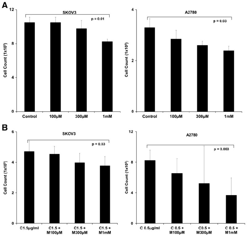
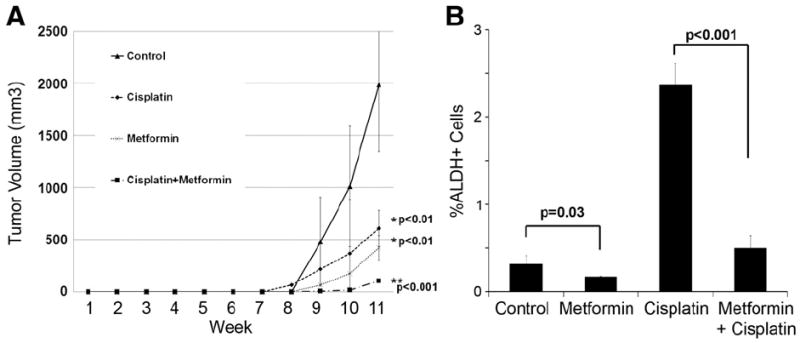
Methods: The impact of metformin on ovarian cancer cell line growth and viability was assessed with trypan blue staining. Aldehyde dehydrogenase (ALDH) expressing CSC were quantified using FACS®. Tumor sphere assays were performed to determine the impact of metformin on cell line and primary human ovarian tumor CSC growth in vitro. In vivo therapeutic efficacy and the anti-CSC effects of metformin were confirmed using both tumor cell lines and ALDH(+) CSC tumor xenografts.

Results: Metformin significantly restricted the growth of ovarian cancer cell lines in vitro. This effect was additive with cisplatin. FACS analysis confirmed that metformin reduced ALDH(+) ovarian CSC. Consistent with this, metformin also inhibited the formation of CSC tumor spheres from both cell lines and patient tumors. In vivo, metformin significantly increased the ability of cisplatin to restrict whole tumor cell line xenografts. In addition, metformin significantly restricted the growth of ALDH(+) CSC xenografts. This was associated with a decrease in ALDH(+) CSC, cellular proliferation, and angiogenesis.

Conclusions: Metformin can restrict the growth and proliferation of ovarian cancer stem cells in vitro and in vivo. This was true in cell lines and in primary human CSC isolates. These results provide a rationale for using metformin to treat ovarian cancer patients.


Conflict of interest statement

Conflict of interest statement

None of the authors have a conflict of interest.

Figures

Fig. 1
Fig. 1
Anti-tumor effects of metformin in vitro. (A) Dose-dependent toxicity of metformin. Cell counts of SKOV3 and A2780 ovarian cancer cells treated with increasing doses of metformin for 3 days. (B) Dose dependent toxicity of metformin when combined with chemotherapy. Cell counts of SKOV3 and A2780 ovarian cancer cells treated with 1.5 μg/mL cisplatin or 0.5 μg/mL of cisplatin, respectively, and metformin (M) 100 μM, 300 μM, and 1 mM for 3 days. For both experiments, cell viability was determined using trypan blue staining. Experiments are representative of 4 independent experiments. All samples were analyzed in triplicate. p values are as indicated.
Fig. 2
Fig. 2
Metformin impacts ovarian CSC. (A1 and 2) FACS analysis of ALDH(+) cells in SKOV3 (1) and A2780 cells (2) after treatment with cisplatin (1.5 μg/mL), metformin (1 mM), or both. DEAB was used as a negative control. Numbers represent the percentage of ALDH(+) cells. (A3 and 4) Average percentage of ALDH(+) cells in untreated controls and cisplatin, metformin, or cisplatin+metformin for SKOV3 (3) and A2780 (4). The results are averages of 4 independent experiments. (B1 and 2) Representative images (200×) of tumor spheres formed from 4000 FACS isolated ALDH(+) SKOV3 cells in the presence or absence of 1 mM metformin and (B2) average sphere numbers. Cells were plated in triplicate and spheres were counted after 14 days. p values are as indicated.
Fig. 3
Fig. 3
Metformin inhibits the formation of tumor spheres in patient ovarian tumor cells. (A) Representative images of patient tumor spheres treated with media or metformin for 14 days. (B) Average number of spheres formed from 4000 ovarian cancer patient ascites cells grown with and without metformin. (C) Average number of spheres formed from 4000 FACS isolated ALDH(+) patient ascites cells grown with and without metformin. All samples were plated in triplicate. p values are as indicated.
Fig. 4
Fig. 4
Metformin restricts ovarian tumor growth in vivo. (A) Tumor growth curves of SKOV3 tumors in mice (i) untreated, or treated (ii) with metformin 150 mg/kg i.p daily, (iii) cisplatin 250 μg/kg i.p for 3 days starting on day 7, or (iv) cisplatin 250 μg/kg i.p. for 3 days starting on day 7+metformin daily starting on day 7 (n=8 animals per treatment group). (B) Kaplan Meier survival curves were calculated for the four treatment groups indicated above. p values are as indicated.
Fig. 5
Fig. 5
Metformin in combination with chemotherapy inhibits proliferation and angiogenesis. (A) Immunohistochemical stain of SKOV3 tumor for CD31 and Ki67 in the indicated treatment groups. (B and C) Quantification of CD31 and Ki67 staining in the four indicated treatment groups. (D) Average percentage of ALDH(+) cells from 4 tumors from each treatment group. p values are as indicated.
Fig. 6
Fig. 6
Metformin restricts the in vivo growth of FACS isolated ALDH(+) ovarian CSC. (A) Tumor growth curves of tumors initiated by FACS isolated ALDH(+) cells and either (i) untreated, or treated with (ii) metformin 150 mg/kg i.p daily, (iii) cisplatin 250 μg/kg i.p for 3 days starting on day 7, or (iv) cisplatin 250 μg/kg i.p. for 3 days starting on day 7+metformin daily (n=8 animals per treatment group). (B) Average percentage of CSC in ALDH(+) cells in representative xenografts from the four treatment groups. p values are indicated.

References

    1. Edwards BK, Brown ML, Wingo PA, Howe HL, Ward E, Ries LA, et al. Annual report to the nation on the status of cancer, 1975–2002, featuring population-based trends in cancer treatment. J Natl Cancer Inst. 2005;97:1407–27. - PubMed
    1. Boman BM, Wicha MS. Cancer stem cells: a step toward the cure. J Clin Oncol. 2008;26:2795–9. - PubMed
    1. Burgos-Ojeda D, Rueda BR, Buckanovich RJ. Ovarian cancer stem cell markers: prognostic and therapeutic implications. Cancer Lett. 2012;322:1–7. - PMC - PubMed
    1. Silva IA, Bai S, McLean K, Yang K, Griffith K, Thomas D, et al. Aldehyde dehydrogenase in combination with CD133 defines angiogenic ovarian cancer stem cells that portend poor patient survival. Cancer Res. 2011;71:3991–4001. - PMC - PubMed
    1. Vazquez-Martin A, Oliveras-Ferraros C, Cufi S, Del Barco S, Martin-Castillo B, Menendez JA. Metformin regulates breast cancer stem cell ontogeny by transcriptional regulation of the epithelial–mesenchymal transition (EMT) status. Cell Cycle. 2010;9:3807–14. - PubMed

Publication types

MeSH terms