. 2012:8:600.
doi: 10.1038/msb.2012.29.

Effects of diet and development on the Drosophila lipidome

Affiliations

Effects of diet and development on the Drosophila lipidome

Maria Carvalho et al. Mol Syst Biol. 2012.

Abstract

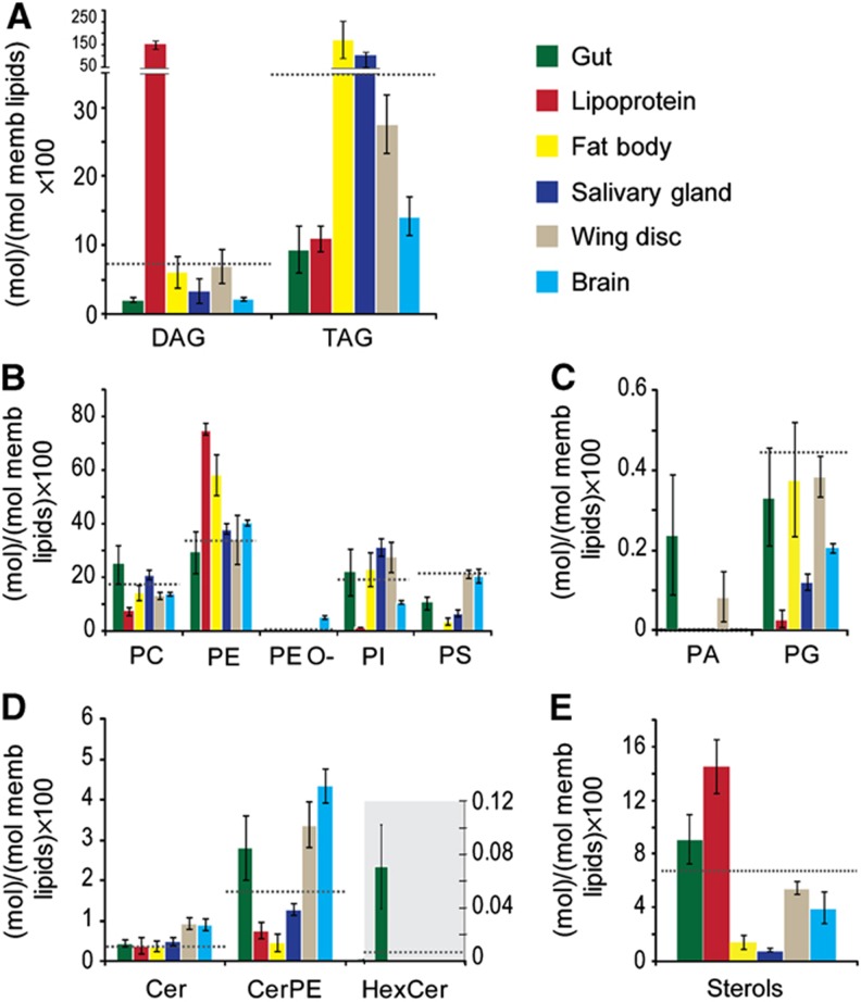
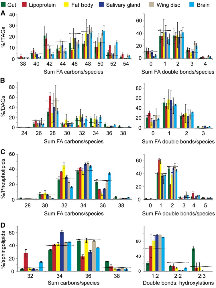
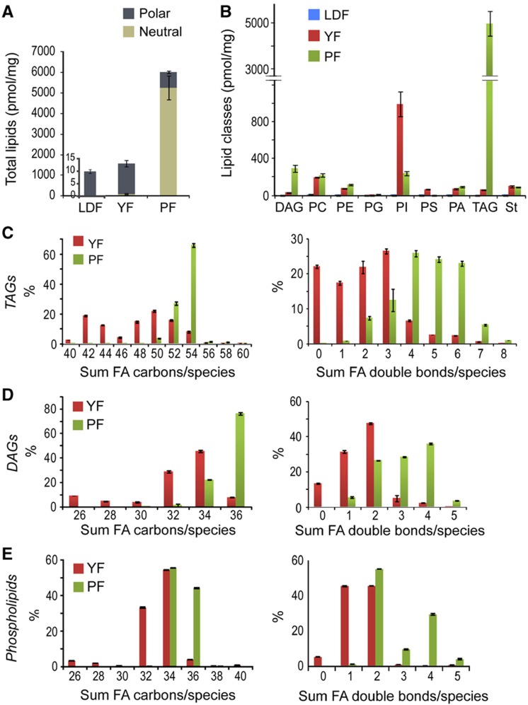
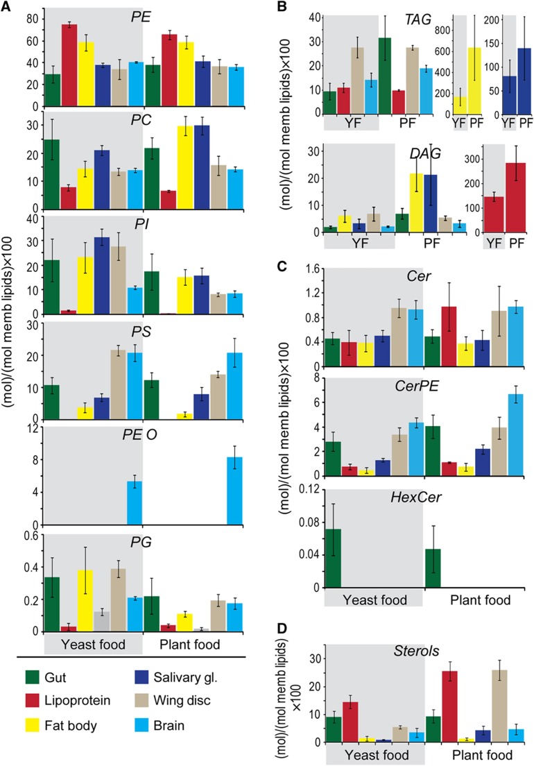
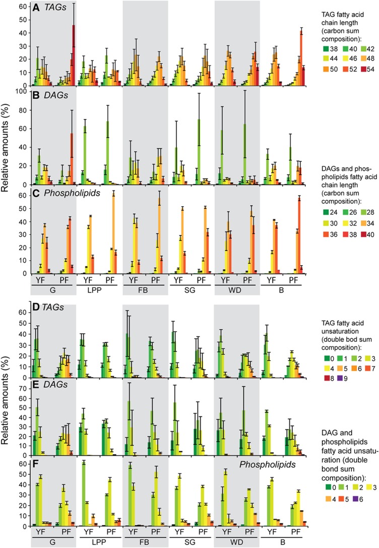
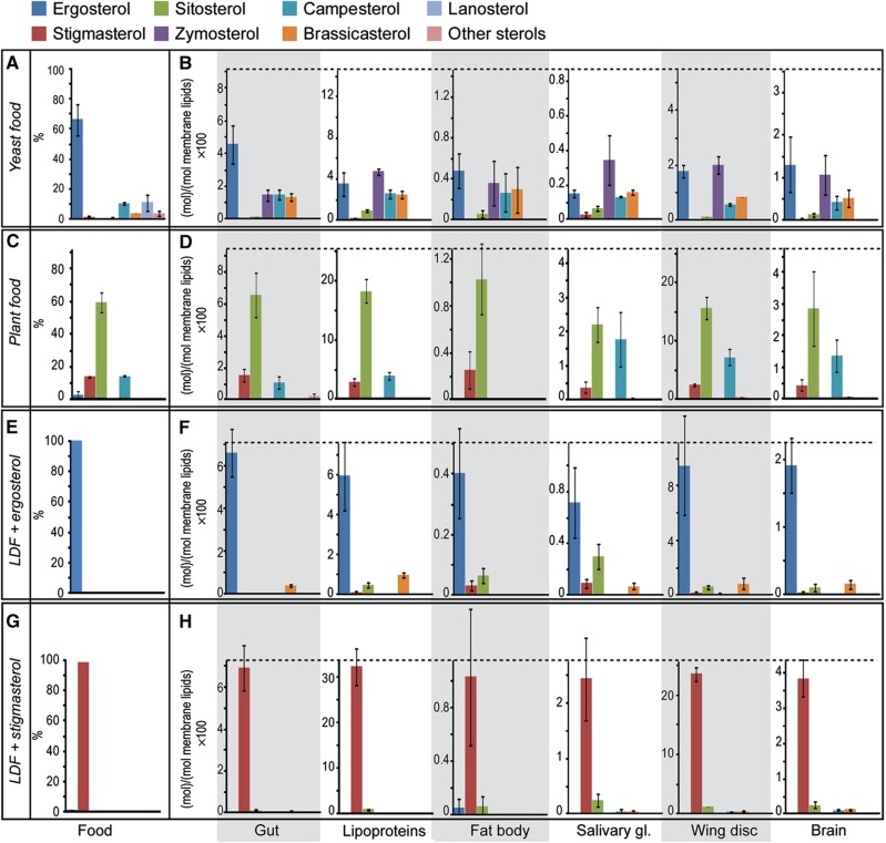
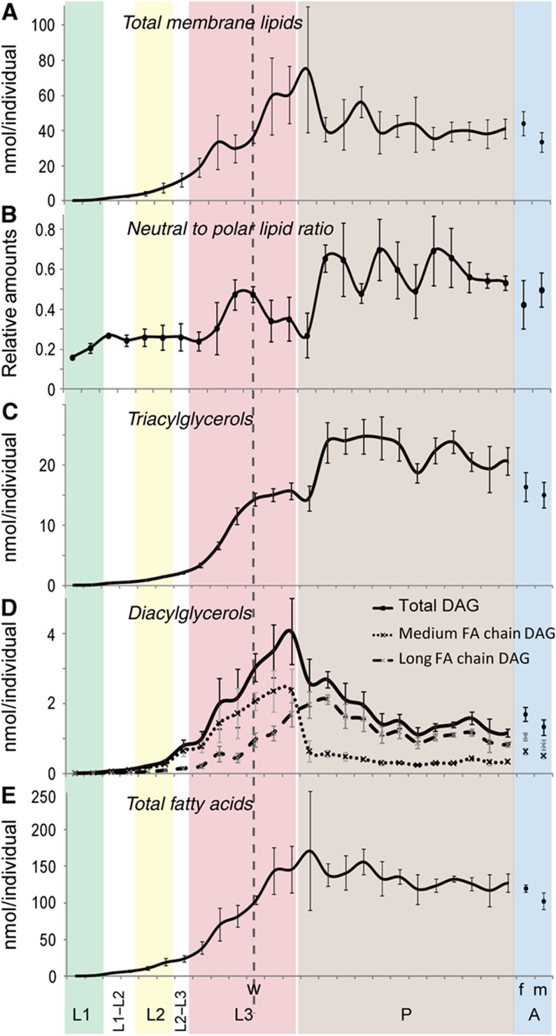
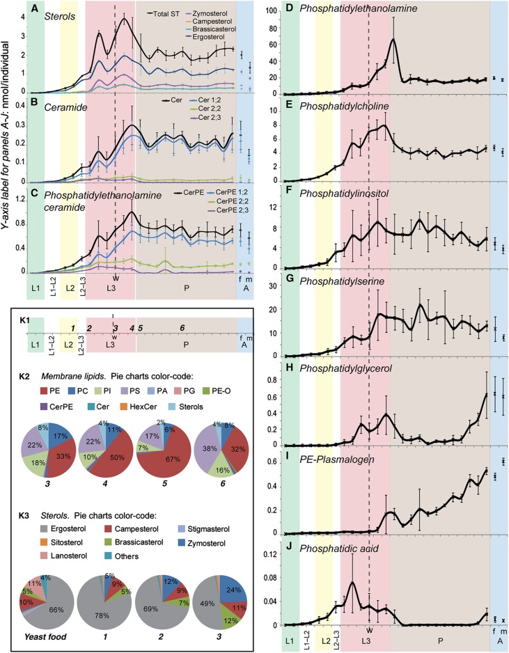
Cells produce tens of thousands of different lipid species, but the importance of this complexity in vivo is unclear. Analysis of individual tissues and cell types has revealed differences in abundance of individual lipid species, but there has been no comprehensive study comparing tissue lipidomes within a single developing organism. Here, we used quantitative shotgun profiling by high-resolution mass spectrometry to determine the absolute (molar) content of 250 species of 14 major lipid classes in 6 tissues of animals at 27 developmental stages raised on 4 different diets. Comparing these lipidomes revealed unexpected insights into lipid metabolism. Surprisingly, the fatty acids present in dietary lipids directly influence tissue phospholipid composition throughout the animal. Furthermore, Drosophila differentially regulates uptake, mobilization and tissue accumulation of specific sterols, and undergoes unsuspected shifts in fat metabolism during larval and pupal development. Finally, we observed striking differences between tissue lipidomes that are conserved between phyla. This study provides a comprehensive, quantitative and expandable resource for further pharmacological and genetic studies of metabolic disorders and molecular mechanisms underlying dietary response.


Conflict of interest statement

The authors declare that they have no conflict of interest.

Figures

Figure 1
Figure 1
Lipid class profiles vary in different tissues. (AE) The abundance of different lipid classes in tissues and lipoproteins of early wandering third instar larvae fed with YF. Each color represents one larval tissue, as indicated. Lipid class abundance is presented as moles per mole of total membrane lipid (phospholipids, sphingolipids and sterols—not including TAG or DAG). (A) Neutral lipids. (B) Major phospholipids. (C) Minor phospholipids. (D) Sphingolipids. (E) Sterols. Error bars indicate standard deviation. Dashed line indicates the lipid class amount in whole larval lipids from animals of the same stage.
Figure 2
Figure 2
Fatty acid chain length and unsaturation varies in different tissues. (AD) The distributions of combined fatty acid chain length (left panels) and unsaturation (right panels) in (A) TAG (B) DAG (C) phospholipids (D) sphingolipids from different tissues of early wandering third instar larvae fed with YF. The abundance of each is presented as a percent relative to all lipids in its category. Dashed line indicates the corresponding profiles of whole larval lipids from animals of the same stage. Error bars indicate standard deviation.
Figure 3
Figure 3
Lipid composition of different Drosophila foods. (A) The amount (pmol/mg) of neutral lipids (brown) and polar lipids (gray) present in LDF, YF and PF. Note that LDF is plotted on a different scale and its lipid content is only 1/150th that of YF. (B) The amount of different lipid classes present in LDF (blue), YF (red) and PF (green). (CE) The relative abundance in YF (red) and PF (green) of lipids with different combined fatty acid chain lengths and unsaturation TAGs (C), DAGs (D) and phospholipids (E) are plotted separately. FA= fatty acid. Error bars indicate standard deviation.
Figure 4
Figure 4
Effect of diet on lipid class abundance in different tissues. (AC) The amount of each lipid class in the different tissues and lipoproteins of early wandering third instar larvae fed either YF (shaded in gray), or PF (not shaded). Tissues are indicated with color, as shown. Lipid class amount is calculated as mol% with respect to total membrane lipids Membrane lipids include phospholipids, sphingolipids and sterols, but not TAG and DAG. (A) Phospholipids (B) Neutral lipids (C) Sphingolipids (D) Sterols. Error bars indicate standard deviation.
Figure 5
Figure 5
Tissue lipids reflect the fatty acids present in the diet. (AF) The distributions of combined fatty acid chain length (A–C) and unsaturation (D–F) in TAG (A, D) DAG (B, E) and phospholipids (C, F) from different tissues of early wandering third instar larvae fed with either YF or PF. Tissues are indicated at the bottom of each chart and are shaded in an alternating pattern to ease comparison: G=gut; LPP=lipoproteins; FB=fat body; SG=salivary gland; WD=wing disk and B=brain. Lipid species with equal fatty acid chain length or unsaturation were pooled together. Phospholipid classes were pooled in one group that represents the average profile of all phospholipids. Length and unsaturation were color coded from green to red according to the color scale as indicated. Green indicates shorter and less saturated fatty acids and red longer and more unsaturated fatty acids. Error bars indicate standard deviation.
Figure 6
Figure 6
Differential uptake, mobilization and tissue accumulation of sterol species. (A, C, E, G) The proportions of different sterols in each food. (B, D, F, H) The amounts of different sterols present in each of the tissues of Drosophila early wandering third instar larvae fed on the corresponding food. The black dashed line indicates the total amount of sterol in each of the tissues. Different sterol species are shown in different colors, as indicated. Sterol amounts are represented as mol% with respect to membrane lipids (including phospholipids, sphingolipids, sterols but not DAG or TAG) (A, B) refer to YF; (C, D) refer to PF; (E, F) refer to LDF+ergosterol and (G, H) refer to LDF+stigmasterol. Error bars indicate standard deviation.
Figure 7
Figure 7
Changes in neutral and polar lipids during development. (AE) The amounts of different types of lipids (nmol/animal) at different developmental stages from hatching to adulthood (time interval=8 h). (A) total membrane lipids, including phospholipids, sphingolipids and sterols, (C) TAG, (D) DAG and (E) total fatty acids. (B) The ratio of neutral lipids (DAGs+TAGs) to polar lipids (membrane lipids) at these different stages. L1=first instar, shaded green; L2=second instar, shaded yellow; L3=third instar, shaded red; P=pupae, shaded brown; A=adults, shaded blue, where m=males and f=females. The dashed line ‘w’ indicates the start of the wandering larval stage. Error bars indicate standard deviation.
Figure 8
Figure 8
Changes in the amounts of different lipid classes during development. (AJ) Amounts (nmol per individual) of different polar lipid classes (indicated) at different developmental stages from hatching to adulthood (time interval=8 h). L1=first instar, shaded green; L2=second instar, shaded yellow; L3=third instar, shaded red; P=pupae, shaded brown; A=adults, shaded blue, where m=males and f=females. The dashed line ‘w’ indicates the start of the wandering larval stage. Error bars indicate standard deviation. (K) Membrane lipid composition at selected developmental time points. (K1) The developmental time points 1–6 that are represented in the pie charts of (K2) and (K3). (K2) Membrane lipids, and highlights the dramatic changes in PE relative to the other membrane lipids that occur during and after pupariation (time points 3–6). (K3) The proportions of different sterol species in YF, and in larvae at time points 1, 2 and 3. It highlights changes in ergosterol and zymosterol accumulation during larval stages.

Similar articles

Cited by

References

    1. Adachi-Yamada T, Gotoh T, Sugimura I, Tateno M, Nishida Y, Onuki T, Date H (1999) De novo synthesis of sphingolipids is required for cell survival by down-regulating c-Jun N-terminal kinase in Drosophila imaginal discs. Mol Cell Biol 19: 7276–7286 - PMC - PubMed
    1. Aldaz S, Escudero LM, Freeman M (2010) Live imaging of Drosophila imaginal disc development. Proc Natl Acad Sci USA 107: 14217–14222 - PMC - PubMed
    1. Baker KD, Thummel CS (2007) Diabetic larvae and obese flies-emerging studies of metabolism in Drosophila. Cell Metab 6: 257–266 - PMC - PubMed
    1. Beadle GW, Tatum EL, Clancy CW (1938) Food level in relation to rate of development and eye pigmentation in Drosophila melanogaster. Biol Bull 75: 447–462
    1. Bernsdorff C, Winter R (2003) Differential properties of the sterols cholesterol, ergosterol, b-sitosterol, trans-7-dehydrocholesterol, stigmasterol and lanosterol on DPPC bilayer order. J Phys Chem B 107: 10658–10664

Publication types

MeSH terms

LinkOut - more resources