. 2012 Sep 13;489(7415):322-5.
doi: 10.1038/nature11317.

Human ES-cell-derived cardiomyocytes electrically couple and suppress arrhythmias in injured hearts

Affiliations

Human ES-cell-derived cardiomyocytes electrically couple and suppress arrhythmias in injured hearts

Yuji Shiba et al. Nature. .

Abstract

Transplantation studies in mice and rats have shown that human embryonic-stem-cell-derived cardiomyocytes (hESC-CMs) can improve the function of infarcted hearts, but two critical issues related to their electrophysiological behaviour in vivo remain unresolved. First, the risk of arrhythmias following hESC-CM transplantation in injured hearts has not been determined. Second, the electromechanical integration of hESC-CMs in injured hearts has not been demonstrated, so it is unclear whether these cells improve contractile function directly through addition of new force-generating units. Here we use a guinea-pig model to show that hESC-CM grafts in injured hearts protect against arrhythmias and can contract synchronously with host muscle. Injured hearts with hESC-CM grafts show improved mechanical function and a significantly reduced incidence of both spontaneous and induced ventricular tachycardia. To assess the activity of hESC-CM grafts in vivo, we transplanted hESC-CMs expressing the genetically encoded calcium sensor, GCaMP3 (refs 4, 5). By correlating the GCaMP3 fluorescent signal with the host ECG, we found that grafts in uninjured hearts have consistent 1:1 host–graft coupling. Grafts in injured hearts are more heterogeneous and typically include both coupled and uncoupled regions. Thus, human myocardial grafts meet physiological criteria for true heart regeneration, providing support for the continued development of hESC-based cardiac therapies for both mechanical and electrical repair.


Figures

Figure 1
Figure 1. Transplanted hESC-CMs partially remuscularize injured guinea pig hearts, preserve mechanical function, and reduce arrhythmia susceptibility
a–b, 28 day-old hESC-CM grafts in a cryoinjured heart stained with picrosirius red (a) and anti-β-myosin heavy chain (βMHC, red) plus human-specific in situ probe (HumCent, brown) (b). c–d, Confocal image of host-graft contact dual-labeled for HumCent (white) and βMHC (red), destained and then immunostained for βMHC (red) and either connexin-43 (e&g, Cx43, green) or cadherins (f&h, green). Arrows indicate Cx43 and cadherin shared between graft and host myocytes. i, LV fractional shortening (FS) by echocardiography in uninjured animals and cryoinjured recipients of PSC vehicle-only, non-CMs or hESC-CMs. j, Representative telemetric ECGs, including non-sustained VT in a hESC-CM recipient (upper), as well as sustained VT and triplet PVCs in a non-CM recipient (lower). k, Percentage of animals by group that showed spontaneous VT during monitoring from days 3–28 post-transplantation. l, Frequency of spontaneous VT by group. m, ECG (red) and stimulation (blue) traces from a cryoinjured non-CM recipient induced to sustained VT by PES. n, Percentage of animals in each group induced to VT. All data are presented as mean ± SEM; n≥13 per group. NS, no significant difference. *, p<0.05 between groups. **, p<0.01 between groups.#, p<0.05 versus day −2.
Figure 2
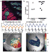
Figure 2. hESC-CM grafts in uninjured hearts show 1:1 coupling with host myocardium
a, GCaMP3+ hESC-CM graft in an uninjured heart immunostained for GFP (green) and βMHC (red), showing extensive host-graft contact with minimal intervening scar. b, Left: GCaMP3+ hESC-CM graft in an uninjured heart during systole and diastole, acquired using an open-chest preparation at 14-days post-transplantation. Right: GCaMP3 fluorescence intensity versus time for the red, green and blue regions of interest, as well as the host ECG (black). GCaMP3 fluorescent transients in all three regions occurred in a 1:1 correspondence with QRS complexes of the host ECG. c, GCaMP3 fluorescent signal (red) and ECG (black) from a representative hESC-CM graft in an uninjured heart, imaged ex vivo under mechanical arrest. 1:1 host-graft coupling occurred during spontaneous beating and pacing at rates ≤5 Hz. Arrow = onset of pacing.
Figure 3
Figure 3. hESC-CM grafts show 1:1 host-graft coupling in a majority of injured recipient hearts, but the extent of coupling and pattern of activation is more variable
a, GCaMP3+ hESC-CM graft in a cryoinjured heart immunostained for GFP (green) and βMHC (red). Small graft nests were located in host muscle within the border zone, but most of the graft was in scar. b, Percentage of visible GCaMP3+ hESC-CM graft in each cryoinjured heart that showed 1:1 host-graft coupling at 14- and 28-days post-transplantation (n=7 and n=15 animals, respectively). c, Representative 28-day-old GCaMP3+ hESC-CM graft in a cryoinjured heart imaged ex vivo during mechanical arrest with blebbistatin and pacing at 3 Hz. Upper: Traces of mean fluorescent intensity versus time for graft regions located within host muscle (‘1’, blue) and the cryoinjury zone (‘2’, red). Both were activated in a 1:1 correlation with the host ECG (black). Lower: Corresponding activation map showing the interval (in ms) between the stimulus pulse and the local rise in GCaMP3 fluorescence. Graft in host muscle (1) showed uniformly rapid activation, while graft in scar (2) activated first in central scar and then gradually progressed toward the border zone. In other instances, graft activation started at the border zone and radiated into the scar. d, GCaMP3 fluorescence and EGG traces (upper) as well as the activation map (lower) for a representative GCaMP3+ hESC-CM graft in a blebbistatin-arrested uninjured heart. This graft showed 1:1 host-graft coupling and a brief interval between stimulus and GCaMP3 transient upstroke.

References

    1. Caspi O, et al. Transplantation of human embryonic stem cell-derived cardiomyocytes improves myocardial performance in infarcted rat hearts. J Am Coll Cardiol. 2007;50:1884–1893. - PubMed
    1. Laflamme MA, et al. Cardiomyocytes derived from human embryonic stem cells in pro-survival factors enhance function of infarcted rat hearts. Nat Biotechnol. 2007;25:1015–1024. - PubMed
    1. van Laake LW, et al. Human embryonic stem cell-derived cardiomyocytes survive and mature in the mouse heart and transiently improve function after myocardial infarction. Stem Cell Research. 2007;1:9–24. - PubMed
    1. Tallini YN, et al. Imaging cellular signals in the heart in vivo: Cardiac expression of the high-signal Ca2+ indicator GCaMP2. Proc Natl Acad Sci U S A. 2006;103:4753–4758. - PMC - PubMed
    1. Tian L, et al. Imaging neural activity in worms, flies and mice with improved GCaMP calcium indicators. Nature Methods. 2009;6:875–881. - PMC - PubMed

Publication types

MeSH terms