. 2012 Oct;464(4):353-65.
doi: 10.1007/s00424-012-1138-3. Epub 2012 Aug 5.

Proteolytic activation of the epithelial sodium channel (ENaC) by the cysteine protease cathepsin-S

Affiliations

Proteolytic activation of the epithelial sodium channel (ENaC) by the cysteine protease cathepsin-S

Silke Haerteis et al. Pflugers Arch. 2012 Oct.

Abstract

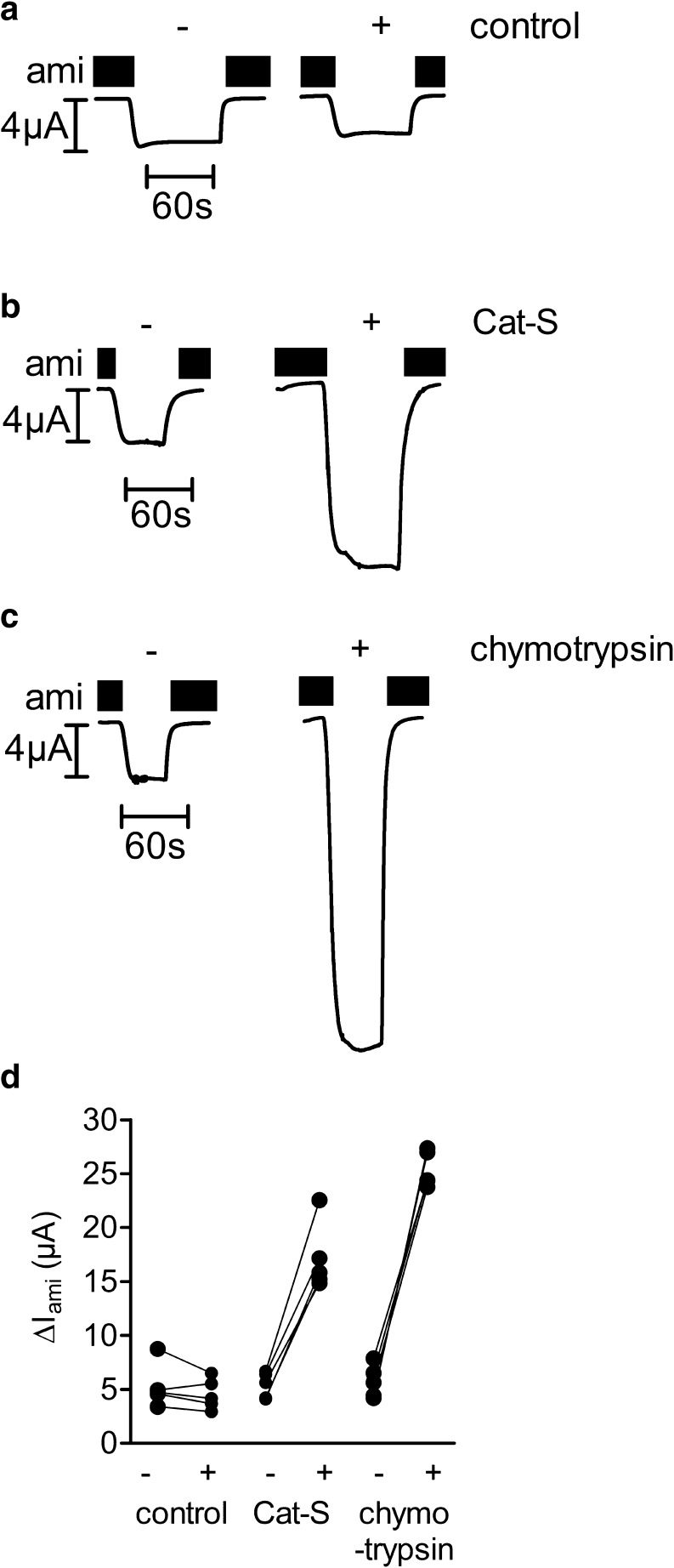
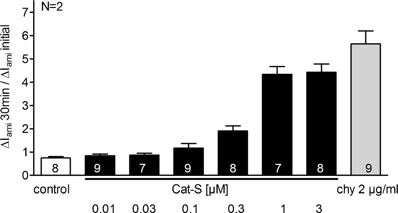
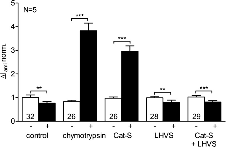
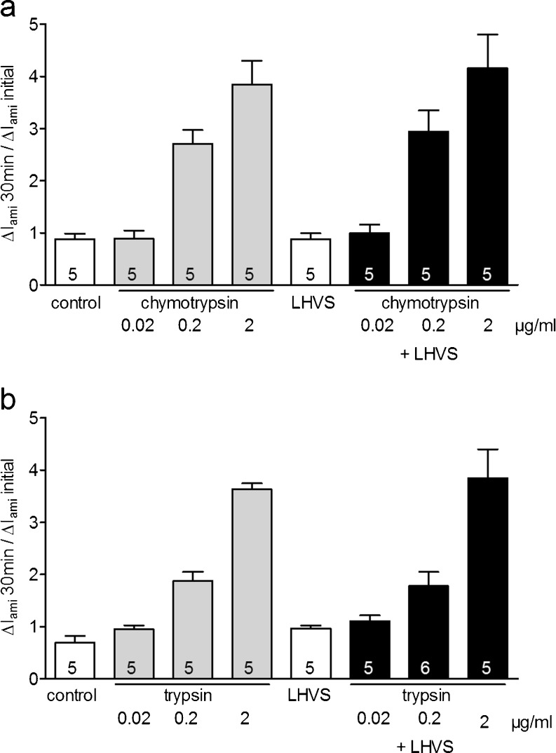
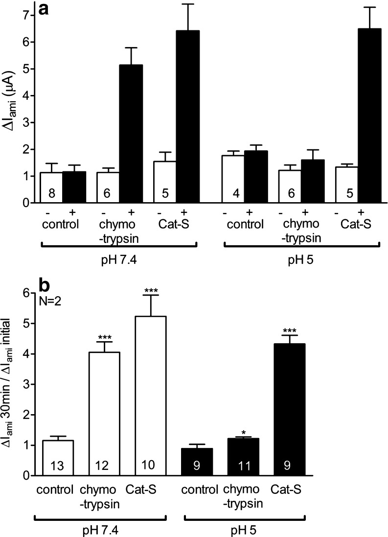
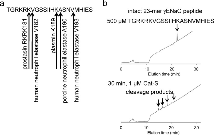
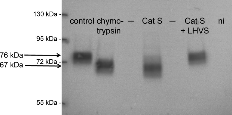
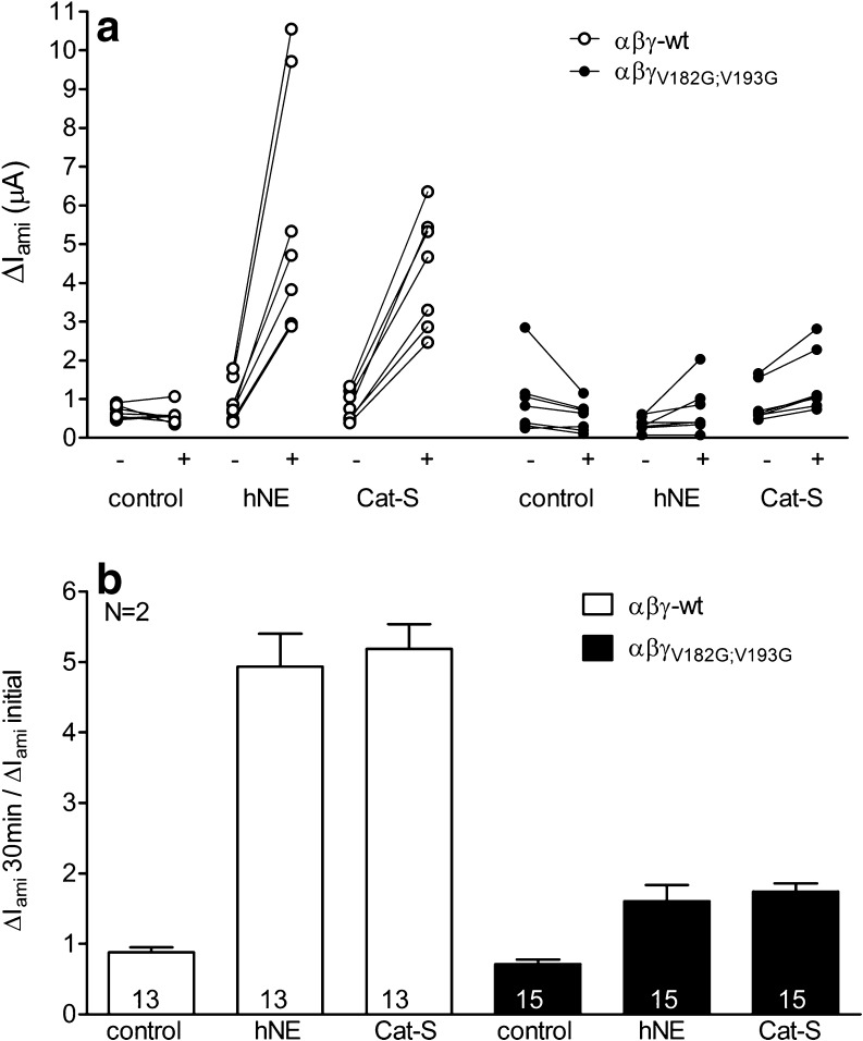
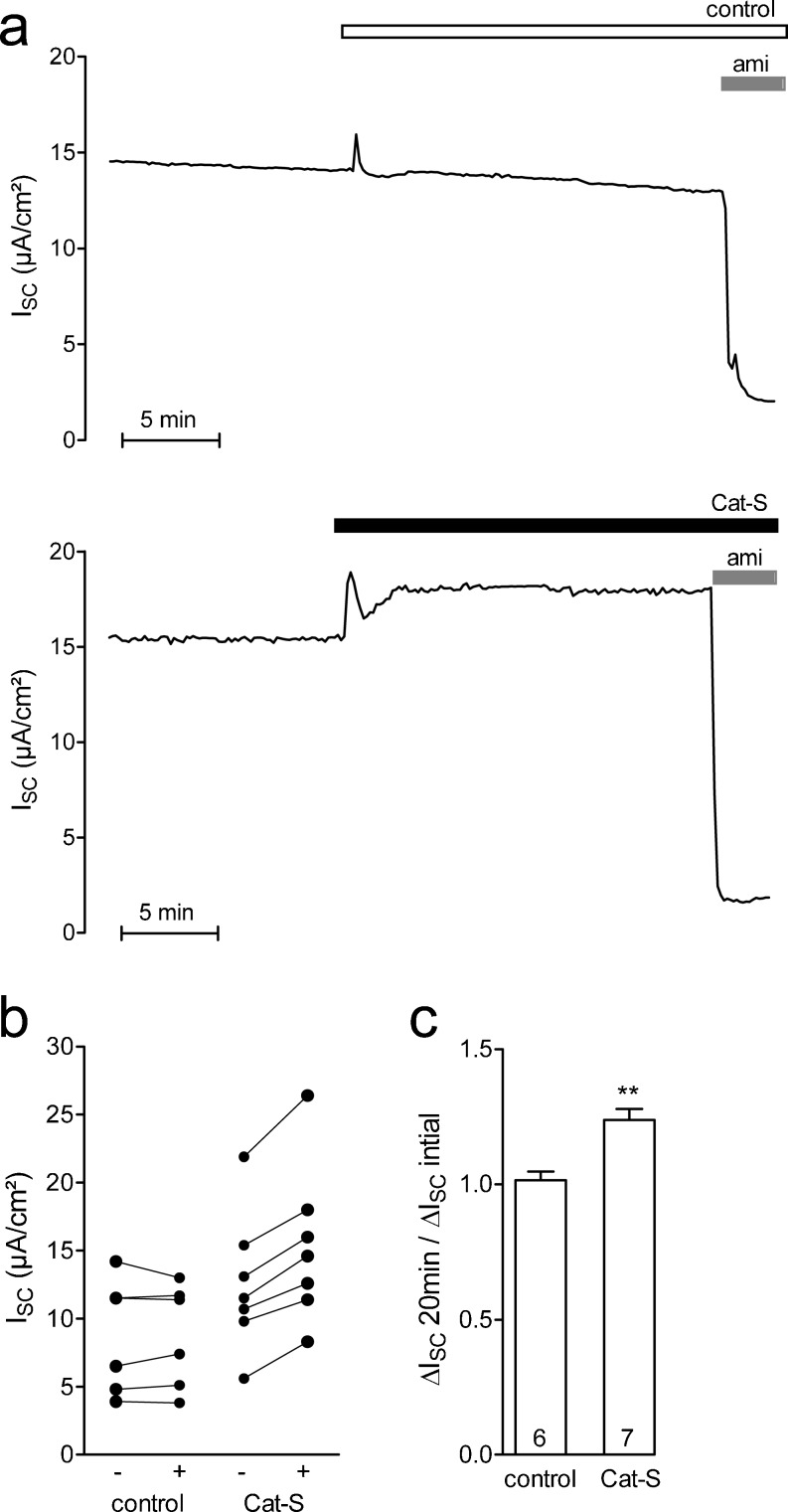
Proteolytic processing of the amiloride-sensitive epithelial sodium channel (ENaC) by serine proteases is known to be important for channel activation. Inappropriate ENaC activation by proteases may contribute to the pathophysiology of cystic fibrosis and could be involved in sodium retention and the pathogenesis of arterial hypertension in the context of renal disease. We hypothesized that in addition to serine proteases, cathepsin proteases may activate ENaC. Cathepsin proteases belong to the group of cysteine proteases and play a pathophysiological role in inflammatory diseases. Under pathophysiological conditions, cathepsin-S (Cat-S) may reach ENaC in the apical membrane of epithelial cells. The aim of this study was to investigate the effect of purified Cat-S on human ENaC heterologously expressed in Xenopus laevis oocytes and on ENaC-mediated sodium transport in cultured M-1 mouse renal collecting duct cells. We demonstrated that Cat-S activates amiloride-sensitive whole-cell currents in ENaC-expressing oocytes. The stimulatory effect of Cat-S was preserved at pH 5. ENaC stimulation by Cat-S was associated with the appearance of a γENaC cleavage fragment at the plasma membrane indicating proteolytic channel activation. Mutating two valine residues (V182 and V193) in the critical region of γENaC prevented proteolytic activation of ENaC by Cat-S. Pre-incubation of the oocytes with the Cat-S inhibitor morpholinurea-leucine-homophenylalanine-vinylsulfone-phenyl (LHVS) prevented the stimulatory effect of Cat-S on ENaC. In contrast, LHVS had no effect on ENaC activation by the prototypical serine proteases trypsin and chymotrypsin. Cat-S also stimulated ENaC in differentiated renal epithelial cells. These findings demonstrate that the cysteine protease Cat-S can activate ENaC which may be relevant under pathophysiological conditions.


Figures

Fig. 1
Fig. 1
Cat-S stimulates ENaC currents in Xenopus laevis oocytes expressing human ENaC. ad Oocytes expressing human ENaC were incubated for 30 min in protease-free solution (control) or in a solution containing either Cat-S (1 μM) or chymotrypsin (2 μg/ml). Amiloride-sensitive whole-cell currents (ΔI ami) were determined before (−) and after (+) incubation. Six representative whole-cell current traces from one batch of oocytes are shown. ac Amiloride (ami) was present in the bath solution to specifically inhibit ENaC as indicated by black bars. d Individual ΔI ami values from a representative experiment using one batch of oocytes. Data points obtained from an individual oocyte are connected by a line
Fig. 2
Fig. 2
Stimulatory effect of Cat-S on human ENaC is concentration dependent. Oocytes expressing human αβγ ENaC were incubated for 30 min in protease-free solution (control), in solutions containing different concentrations of Cat-S (0.01, 0.03, 0.1, 0.3, 1, and 3 μM) or in a solution containing chymotrypsin (2 μg/ml). Amiloride-sensitive whole-cell currents (ΔI ami) were detected before (ΔI ami initial) and after incubation (ΔI ami 30 min). Columns represent the relative stimulatory effect on ΔI ami calculated as the ratio of ΔI ami 30 min/ΔI ami initial. Numbers inside the columns indicate the number of individual oocytes measured. N indicates the number of different batches of oocytes
Fig. 3
Fig. 3
Activation of ENaC by Cat-S is prevented by the Cat-S inhibitor LHVS. Oocytes expressing human ENaC were incubated for 30 min in protease-free solution (control), in chymotrypsin (2 μg/ml), in Cat-S (1 μM), in LHVS (2 μM), or in a solution containing a combination of Cat-S (1 μM) and LHVS (5 μM). Amiloride-sensitive whole-cell currents (ΔI ami) were determined before (−) and after (+) incubation. The bar diagram represents normalized average results obtained in five different batches of oocytes (N = 5). The individual ΔI ami values were normalized to the mean ΔI ami value of the ENaC-expressing control group in protease-free solution. Numbers inside the columns indicate the number of individual oocytes measured. **p < 0.01, ***p < 0.001, paired t test
Fig. 4
Fig. 4
The Cat-S inhibitor LHVS has no effect on ENaC activation by the serine proteases trypsin and chymotrypsin. a Oocytes expressing human ENaC were incubated for 30 min in protease-free solution (control), in solutions with different concentrations of chymotrypsin (0.02, 0.2, and 2 μg/ml), in LHVS (2 μM), or in solutions containing a combination of different concentrations of chymotrypsin (0.02, 0.2, and 2 μg/ml) and LHVS (2 μM). Amiloride-sensitive whole-cell currents (ΔI ami) were determined before (−) and after (+) incubation. Columns represent relative stimulatory effect on ΔI ami calculated as the ratio of ΔI ami measured after 30 min of incubation (ΔI ami 30 min) to the initial ΔI amiI ami initial) measured before incubation. Numbers inside the columns indicate the number of individual oocytes measured. b Similar experiment as shown in a using the serine protease trypsin (2 μg/ml) instead of chymotrypsin
Fig. 5
Fig. 5
Cat-S can activate ENaC in an acidic environment. Oocytes expressing human ENaC were incubated for 30 min in protease-free (control), in chymotrypsin (0.2 μg/ml), or in Cat-S (1 μM) solution with a physiological pH of 7.4 (white columns) or an acidic pH of 5 (black columns). Amiloride-sensitive whole-cell currents (ΔI ami) were determined before (−) and after (+) incubation. a ΔI ami values from a representative experiment using one batch of oocytes. b Summary of similar experiments as shown in a. Columns represent relative stimulatory effect on ΔI ami calculated as the ratio of ΔI ami measured after 30 min of incubation (ΔI ami 30 min) to the initial ΔI amiI ami initial) measured before incubation. Numbers inside the columns indicate the number of individual oocytes measured. N indicates the number of different batches of oocytes. *p < 0.05, ***p < 0.001, unpaired t test
Fig. 6
Fig. 6
In vitro cleavage analysis of the 23-mer γENaC peptide suggests that Cat-S may cleave human γENaC at more than one cleavage site. a Sequence of the 23-mer γENaC peptide showing putative cleavage sites for proteolytic ENaC activation. b The 23-mer γENaC peptide (500 μM) was incubated with Cat-S (1 μM) for 30 min and cleavage products were identified by HPLC and mass spectrometry. Cat-S degraded the 23-mer γENaC peptide showing four products detected by HPLC
Fig. 7
Fig. 7
Activation of ENaC by Cat-S generates a γENaC cleavage product at the cell surface indicating proteolytic channel activation. The Cat-S inhibitor LHVS prevented the generation of an additional cleavage product at the cell surface. Oocytes expressing human ENaC were incubated for 30 min in protease-free solution (control), in chymotrypsin (2 μg/ml), in Cat-S (1 μM), or in a solution containing a combination of Cat-S (1 μM) and LHVS (5 μM). Expression of biotinylated γENaC at the cell surface was analyzed by SDS-PAGE. γENaC was detected with an antibody against the C-terminus of human γENaC. In non-injected (ni) oocytes, γENaC-specific signals were absent. “–” indicates an empty lane on the gel. Molecular weight markers are shown on the left side of the gel. Representative western blot from one batch of oocytes
Fig. 8
Fig. 8
Mutating two putative neutrophil elastase cleavage sites (γV182;V193) prevents proteolytic activation of γENaC by Cat-S. Oocytes expressing αβγ (open symbols) or αβγV182G;V193GENaC (filled symbols) were incubated for 30 min in protease-free solution (control) or in a solution containing either hNE (10 μg/ml) or Cat-S (1 μM). Amiloride-sensitive whole-cell currents (ΔI ami) were determined before (−) and after (+) incubation. a Individual ΔI ami values from a representative experiment using one batch of oocytes. Data points obtained from individual oocytes are connected by a line. b Summary of similar experiments as shown in a. Columns represent relative stimulatory effect on ΔI ami calculated as the ratio of ΔI ami measured after 30 min of incubation (ΔI ami 30 min) to the initial ΔI amiI ami initial) measured before incubation. Numbers inside the columns indicate the number of individual oocytes measured. N indicates the number of different batches of oocytes
Fig. 9
Fig. 9
Cat-S stimulates ENaC in confluent M-1 mouse collecting duct cells. a Representative equivalent short-circuit current (I SC) recordings from confluent M-1 cells pretreated with nafamostate mesylate to reduce constitutive ENaC activation by endogenous proteases. Vehicle control (phosphate buffered saline, upper trace) or Cat-S (2 μM, lower trace) was added to the apical bath solution of M-1 cells. At the end of the experiment, amiloride (ami; 10 μM) was added apically to confirm that the stimulated I SC was mediated by ENaC. b, c Summary of results from similar experiments as shown in a. b Data points represent individual I SC values obtained from six (control) or seven (Cat-S) individual experiments that are connected by a line. c Columns represent relative stimulatory effect on ΔI SC calculated as the ratio of ΔI SC measured after 20 min of incubation (ΔI SC 20 min) to the initial I SCI SC initial) measured before apical addition. **p < 0.01, unpaired t test

References

    1. Adebamiro A, Cheng Y, Rao US, Danahay H, Bridges RJ. A segment of γENaC mediates elastase activation of Na+ transport. J Gen Physiol. 2007;130:611–629. doi: 10.1085/jgp.200709781. - DOI - PMC - PubMed
    1. Ayesa S, Lindquist C, Agback T, Benkestock K, Classon B, Henderson I, Hewitt E, Jansson K, Kallin A, Sheppard D, Samuelsson B. Solid-phase parallel synthesis and SAR of 4-amidofuran-3-one inhibitors of cathepsin S: effect of sulfonamides P3 substituents on potency and selectivity. Bioorg Med Chem. 2009;17:1307–1324. doi: 10.1016/j.bmc.2008.12.020. - DOI - PubMed
    1. Bengrine A, Li J, Hamm LL, Awayda MS. Indirect activation of the epithelial Na+ channel by trypsin. J Biol Chem. 2007;282:26884–26896. doi: 10.1074/jbc.M611829200. - DOI - PubMed
    1. Bertog M, Letz B, Kong W, Steinhoff M, Higgins MA, Bielfeld Ackermann A, Frömter E, Bunnett NW, Korbmacher C. Basolateral proteinase-activated receptor (PAR-2) induces chloride secretion in M-1 mouse renal cortical collecting duct cells. J Physiol. 1999;521:3–17. doi: 10.1111/j.1469-7793.1999.00003.x. - DOI - PMC - PubMed
    1. Brix K, Dunkhorst A, Mayer K, Jordans S. Cysteine cathepsins: cellular roadmap to different functions. Biochimie. 2008;90:194–207. doi: 10.1016/j.biochi.2007.07.024. - DOI - PubMed

Publication types

MeSH terms