Review
. 2013 Feb;70(4):661-87.
doi: 10.1007/s00018-012-1073-7. Epub 2012 Aug 3.

Aurora A kinase (AURKA) in normal and pathological cell division

Affiliations
Review

Aurora A kinase (AURKA) in normal and pathological cell division

Anna S Nikonova et al. Cell Mol Life Sci. 2013 Feb.

Abstract

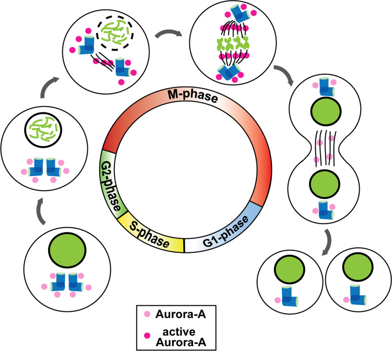
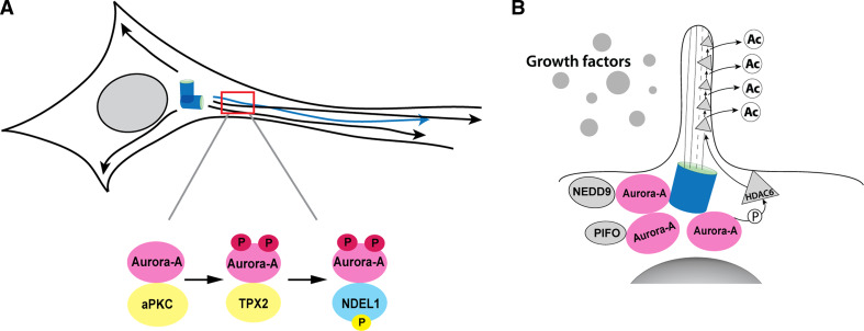
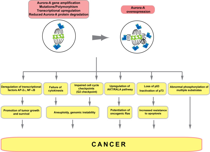
Temporally and spatially controlled activation of the Aurora A kinase (AURKA) regulates centrosome maturation, entry into mitosis, formation and function of the bipolar spindle, and cytokinesis. Genetic amplification and mRNA and protein overexpression of Aurora A are common in many types of solid tumor, and associated with aneuploidy, supernumerary centrosomes, defective mitotic spindles, and resistance to apoptosis. These properties have led Aurora A to be considered a high-value target for development of cancer therapeutics, with multiple agents currently in early-phase clinical trials. More recently, identification of additional, non-mitotic functions and means of activation of Aurora A during interphase neurite elongation and ciliary resorption have significantly expanded our understanding of its function, and may offer insights into the clinical performance of Aurora A inhibitors. Here we review the mitotic and non-mitotic functions of Aurora A, discuss Aurora A regulation in the context of protein structural information, and evaluate progress in understanding and inhibiting Aurora A in cancer.


Figures

Fig. 1
Fig. 1
Aurora A in the cell cycle. Aurora A begins to accumulate significantly at centrosomes in the S phase and is activated at the boundary between the G2 and M phases. Active Aurora A propagates along the mitotic spindle to the midzone, with most of the protein being inactivated and degraded before cytokinesis with only low levels detectable in early G1 cells
Fig. 2
Fig. 2
Aurora A interaction with partners regulating activation and destruction. Proteins interacting with Aurora to promote activation (green line, star), promote destruction (blue line, arrow), or protect from destruction (orange line, shield) are indicated in context of phases of cell cycle in which they associate with Aurora A. When interaction involves specific phosphorylations on Aurora A, these are indicated in a circle at the left of drawing
Fig. 3
Fig. 3
Active Aurora A in non-cycling cells. a aPKC activates Aurora A, which interacts with NDEL1 to induce microtubule-based extension of post-mitotic neurons. b NEDD9 and PIFO activate Aurora A at the basal body of cilia in quiescent cells, leading to Aurora-A-dependent activation of HDAC6, and ciliary resorption
Fig. 4
Fig. 4
a Ribbon diagram of the ADP-bound structure of human Aurora A from PDB entry 1OL5. The chain is colored from blue (N-terminus) to red (C-terminus). The activation loop is shown in magenta and the activator TPX2 is colored beige. Aurora A in this structure is phosphorylated on T287 and T288, and the DFG motif at the beginning of the activation loop is in the “DFG-in” position, such that the F275 of the DFG motif (magenta sticks) is located under the C-helix (cyan). b Close-up of the active site of Aurora A from PDB entry 1OL5. Hydrogen bonds are shown in dotted black lines between pT288 and R255 of the HRD motif, T292 of the activation loop and D256 of the HRD motif and K258. The yellow spheres are active site magnesium ions. The regulatory spine of Aurora A is shown in dots (H254, F275, Q195, and L196). c Several structures in the “DFGup” conformation are shown. F275 points upwards between the C-helix and the N-terminal domain β sheet. d Compound MLN8054 bound to human Aurora A from PDB entry 2WTV. F275 of the DFG motif is in a “DFGup” position, disrupting the salt-bridge interaction between E181 and K162. The activation loop is in an inactive position, in contrast to the active position in a and b
Fig. 5
Fig. 5
Aurora A activities in cancer. Aurora A expression is elevated based on changes in DNA copy number, transcription, and/or changes in protein stability. Overexpressed Aurora A induces multiple categories of growth defect that promote cancer, outlined here schematically; see text for details
Fig. 6
Fig. 6
Extended Aurora A interaction network. Schematic indicates proteins that known or highly likely to interact physically and/or functionally (indirect interaction) with Aurora A/AURKA. All proteins are indicated by official gene symbol. Interaction data were collected using online databases String [251] (using medium confidence score 0.4 and excluding text-mining-only results) and Ingenuity [http://www.ingenuity.com/] (extracting only experimental and high confidence predicted interactions). Data were imported, merged, and visualized in Cytoscape [252]. Blue rim around protein indicates direct protein–protein interaction; lack of rim indicates indirect interaction. Green circles represent highly validated interactions generally reported in publications. Blue circles represent high confidence hits from larger screens, but are less well studied. Green lines indicate interactions with Aurora A/AURKA; blue lines indicate interactions among proteins described as interacting with Aurora A/AURKA

References

    1. Chan CS, Botstein D. Isolation and characterization of chromosome-gain and increase-in-ploidy mutants in yeast. Genetics. 1993;135:677–691. - PMC - PubMed
    1. Paris J, Philippe M. Poly(A) metabolism and polysomal recruitment of maternal mRNAs during early Xenopus development. Dev Biol. 1990;140:221–224. doi: 10.1016/0012-1606(90)90070-Y. - DOI - PubMed
    1. Andresson T, Ruderman JV. The kinase Eg2 is a component of the Xenopus oocyte progesterone-activated signaling pathway. EMBO J. 1998;17:5627–5637. doi: 10.1093/emboj/17.19.5627. - DOI - PMC - PubMed
    1. Roghi C, Giet R, Uzbekov R, Morin N, Chartrain I, Le Guellec R, Couturier A, Doree M, Philippe M, Prigent C. The Xenopus protein kinase pEg2 associates with the centrosome in a cell cycle-dependent manner, binds to the spindle microtubules and is involved in bipolar mitotic spindle assembly. J Cell Sci. 1998;111(Pt 5):557–572. - PubMed
    1. Glover DM, Leibowitz MH, McLean DA, Parry H. Mutations in aurora prevent centrosome separation leading to the formation of monopolar spindles. Cell. 1995;81:95–105. doi: 10.1016/0092-8674(95)90374-7. - DOI - PubMed

Publication types

MeSH terms

LinkOut - more resources